Categories:

Temperature and Pulse Basics & Monthly Log

Also see:
Ray Peat, PhD on Thyroid, Temperature, Pulse, and TSH
Low Carb Diet – Death to Metabolism
Body Temperature, Metabolism, and Obesity
Thyroid, Temperature, Pulse
Metabolism, Brain Size, and Lifespan in Mammals
Promoters of Efficient v. Inefficient Metabolism
Inflammation from Decrease in Body Temperature
Melatonin Lowers Body Temperature
Menopausal Estrogen Therapy Lowers Body Temperature
Thyroid Function, Pulse Rate, & Temperature
“Curing” a High Metabolic Rate with Unsaturated Fats
Fat Deficient Animals – Activity of Cytochrome Oxidase
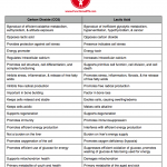
Comparison: Carbon Dioxide v. Lactic Acid
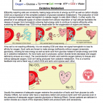
Carbon Dioxide Basics
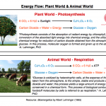
Energy Flow: Plant World and Animal World
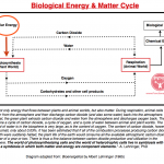
Biological Energy & Matter Cycle
Is 98.6 Really Normal?

Metabolism is the sum of chemical processes that occur in an organism in order to maintain life. Life depends upon the continual conversion of fuel substrates into chemical energy, allowing cells to perform biological work. Heat is produced by these cellular metabolic processes so the resting metabolic rate can be predicted accurately by the rate of heat production.

The unifying principle of Ray Peat, PhD’s work in FPS’ opinion is that energy production from cellular respiration/metabolism allows for structure and function of cells to be optimized, and this improved structure and function promotes continued high energy output as well as the production of protective steroid hormones and carbon dioxide.

Anything that interferes with energy production has opposing effects, slowing energy output, the consumption of oxygen, and the production of protective hormones and carbon dioxide. The ability to produce energy is at the center of health v. non health, youth v. aging, etc.

A simple way to monitor the intensity of your resting metabolism (i.e. how well you are making energy/heat) is to track the resting oral temperature and pulse rate. Collect this data upon waking while lying in bed, ~40 minutes after breakfast, and between 1 and 3 pm in the afternoon.

Prior to 1940, the resting body temperature upon waking was a common way in which physicians would diagnose a slow metabolism. If the temperature was below optimal, a trial of natural desiccated thyroid was given. If symptoms regressed as metabolic efficiency improved and temperature rose from the thyroid supplementation, the therapy was continued. Broda Barnes, MD, PhD and other doctors influenced by his work use this method during his career with much success.

Here are some temperature and pulse tracking basics:
1. Before taking each reading, be at rest for at least five minutes. Use a basal thermometer or mercury thermometer for oral temperature accuracy. An oximeter (like one from Santa Medical) can help you quickly track your pulse. If you don’t have an oximeter, count your heart beats at the neck or wrist for a full 60 seconds or count for ten seconds and multiply by six.
2. Temperature or pulse should not decrease following meals. If it does this consistently, adrenal stress hormones are playing a significant role in your physiology. The introduction of sugar from food lowers the stress hormones and provides a more clear outlook on the resting metabolism.

“If the night-time stress is very high, the adrenalin will still be high until breakfast, increasing both temperature and pulse rate. The cortisol stimulates the breakdown of muscle tissue and its conversion to energy, so it is thermogenic, for some of the same reasons that food is thermogenic.

After eating breakfast, the cortisol (and adrenalin, if it stayed high despite the increased cortisol) will start returning to a more normal, lower level, as the blood sugar is sustained by food, instead of by the stress hormones. In some hypothyroid people, this is a good time to measure the temperature and pulse rate. In a normal person, both temperature and pulse rate rise after breakfast, but in very hypothyroid people either, or both, might fall.” -Ray Peat, PhD

3. Upon waking, an ideal temperature is between 97.8-98.6F and a pulse rate between 75-85 beats per minute (BPM). Other readings during the day should fall within these parameters as well with temperatures being closer to 98.6F than 97.8F. Data points below the optimal are a sign of a slowed metabolic rate.
4. There should be an increase in temperature and pulse rate following a good breakfast as the liver becomes energized allowing it to form the active thyroid hormone, triiodonthyronine (T3).
5. The afternoon temperature and pulse should increase relative to the morning readings because of the thermogenic effect of good nutrition and movement, and the metabolic stimulation from light.
6. In some individuals with overactive adrenal stress hormones (adrenaline/cortisol), the temperature and pulse rate may seem optimal despite symptoms that indicate otherwise. In such people, the temperature and/or pulse will start to drop when metabolic efficiency starts to be restored. This is a sign of progress. Anything consistently over the optimal readings is either a sign of hypermetabolism or an exaggerated adrenaline/cortisol response.
7. Correlate the temperature and pulse rate data to the person. The temperature and pulse information serves as one piece of data that needs context to be understood completely.
8. The ease by which this data can be taken and tracked makes it ideal in discovering which foods, supplements, and activities promote or degrade your metabolism.
9. If you can’t get all three readings due to lifestyle/work commitments during the weekdays, do your best to at least get the waking temperature/pulse and be religious about getting all three readings on the weekend. Setting an alarm or event in your calendar on your phone can serve as a reminder until tracking becomes more habitual.
10. Both the temperature and pulse rate provide more data together than either one of them alone.

Click the link below for a .pdf of a Monthly Temperature and Pulse Log to print and use.
Temperature and Pulse Log by FPS

KEY for the log:
WT/P = Waking Temperature/Pulse
ABT/P = After Breakfast Temperature/Pulse
AT/P = Afternoon Temperature/Pulse

Posted in General.

Tagged with , , , , , , .


Collection of FPS Charts

Also see:
Master List – Ray Peat, PhD Interviews
Collection of Ray Peat Quote Blogs by FPS

Promoters of Efficient v. Inefficient Metabolism

Comparison: Oxidative Metabolism v. Glycolytic Metabolic

Comparison: Carbon Dioxide v. Lactic Acid

Carbon Dioxide Basics

Low Blood Sugar Basics

Temperature and Pulse Basics & Monthly Log

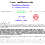
Protect the Mitochondria

Energy Flow: Plant World and Animal World

The Sun: Source of All Biological Energy

Biological Energy & Matter Cycle

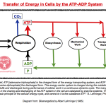
Transfer of Energy in Cells by the ATP-ADP System

Supplement Schedule Chart

FPS Supplement Schedule

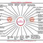
Common Paths to a Low Metabolism

fps-metabolism-chart-fps

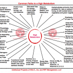
Common Paths to a High Metabolism

FPS High Metabolism

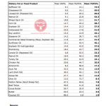
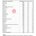
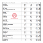
Charts: Mean SFA, MUFA, & PUFA Content of Various Dietary Fats

Sorted by Mean Percentage of Polyunsaturated Fatty Acids (PUFA) - Highest to Lowest

Sorted by Mean Percentage of Polyunsaturated Fatty Acids (PUFA) – Highest to Lowest

Sorted by Mean Percentage of Saturated Fatty Acids (SFA) - Highest to Lowest

Sorted by Mean Percentage of Saturated Fatty Acids (SFA) – Highest to Lowest

Sorted by SFA%:PUFA% Ratio - Highest to Lowest (Most Safe to Least Safe)

Sorted by SFA%:PUFA% Ratio – Highest to Lowest
(Most Safe to Least Safe)

Stress — A Shifting of Resources

fps stress shift

Bodily Resources vs Demands

Posted in General.

Tagged with , , , , .


Low Blood Sugar Basics

Also see:
Ray Peat, PhD on Low Blood Sugar & Stress Reaction
PUFA Promote Stress Response; Saturated Fats Suppress Stress Response
Protect the Mitochondria
Saturated and Monousaturated Fatty Acids Selectively Retained by Fat Cells
PUFA Decrease Cellular Energy Production
The Randle Cycle
Low Carb Diet – Death to Metabolism
Free Fatty Acid Suppress Cellular Respiration
Blood Sugar – Resistance to Allergy and Shock
Sugar (Sucrose) Restrains the Stress Response
Protection from Endotoxin
Possible Indicators of High Cortisol and Adrenaline
Thyroid peroxidase activity is inhibited by amino acids
Toxicity of Stored PUFA
Belly Fat, Cortisol, and Stress
Sugar (Sucrose) Restrains the Stress Response
A long childhood feeds the hungry human brain

“Every stress leaves an indelible scar, and the organism pays for its survival after a stressful situation by becoming a little older.” -Hans Selye

“Since glucose is the main fuel for the brain, and since the human brain is the factor that elevates man above other animals, mother nature took special precautions against a lack of glucose in the bloodstream at all times.” -Broda Barnes & Charlotte Barnes

“In other words, the thyroid has a profound effect on the liver. We have other evidence that a lack of thyroid is accompanied by a sluggish liver…Since a sluggish liver is the most common cause of hypoglycemia, it should follow that the hypothyroid patient is highly susceptible to low blood sugar.” -Broda Barnes, MD, PhD and Charlotte Barnes

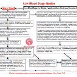
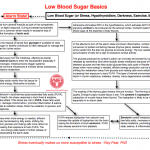
The flow chart thumbnail below is inspired by the work of Ray Peat, PhD and attempts to identify the major players involved in the regulation of blood sugar in the event of hypoglycemia. The alarm or stress reaction that occurs during low blood sugar is a lifesaver during actual starvation or when we can’t get to food, but it should ideally only turn on occasionally.

The more we enter survival (alarm) mode, the greater the pituitary and adrenal activity become dominant relative to thyroid activity. If the blood sugar is chronically low due to illogical food choices or imbalanced or infrequent meals, the alarm state is active too frequently and problems develop over time.

The heightened sympathetic nervous system activity during low blood sugar is like that which happens in hypothyroidism & during prolonged darkness, exercise, and malnutrition. The interconnectedness of the stress mediators is significant since they tend to promote each other in vicious, self-accelerating loops especially when the tissues are rich in polyunsaturates, if the bowel is toxic, or in a backdrop of low carbon dioxide or high estrogen.

The response to stress changes with age in relation to our previous dietary choices. In youth (assuming an adequate diet), the relative deficiency of stored unsaturates, high thyroid, glycogen availability, and optimal protective steroid hormone production produces a self-limiting stress response instead of a self-stimulating one.

As the tissues become more unsaturated with aging or due to poor food choice, the stress response switches from adaptive to dysadaptive, making the body progressively less capable of handling future stresses without producing inflammation and other adverse effects. The unsaturates’ anti-thyroid actions slow the synthesis of protective steroid hormones (pregnenolone, DHEA, progesterone) from cholesterol; cortisol becomes the dominant stress steroid and the ratio of cortisol to testosterone & estrogen to progesterone increases.

Using multiple means, the body protectively slows the metabolism (goes into economy mode) during stress to prolong survival since the body consumes itself during such times. This effect is favorable if you’re actually starving and food isn’t available but not friendly if long-term weight management is desired. Immune function is also suppressed, making the body more susceptible to infection and sickness.

The ability to store enough glycogen to handle stress lessens the need for adrenal activity. With thyroid suppression comes less ability to store glycogen, making low blood sugar and the alarm state more common. Without an energized liver, the conversion of T4 to T3 becomes less efficient, increasing adrenal and pituitary activity. An increased dependence on cortisol to provide glucose for fuel results, wasting protein rich-tissues like skeletal muscle. The loss of muscle tissue (and bone mass) is a characteristic of aging.

As the thyroid activity is suppressed, liver function suffers allowing estrogen to accumulate. Estrogen further blocks thyroid function, depletes glycogen, increases fatty acids, amplifies endotoxin’s effects, is toxic to the liver, and promotes inflammation in another cruel cycle.

Blood sugar balance using protective and digestible food choices is a fundamental of good nutrition practices. While some will argue that we don’t need to eat carbohydrate because our body can make carbohydrate from itself, that side of the fence is looking at physiology through a pin hole and misses the big picture. Ample carbohydrate particularly from ripe fruits, orange juice, milk, honey, and sucrose keeps the alarm state and vicious inflammatory cycles at bay.

The body uses its own tissues to make glucose during hypoglycemia because glucose is important in maintaining optimal function. Without enough dietary carbohydrate, the body becomes dependent on stress hormones for glucose. For optimal health, sustain blood sugar with food, not stress hormones.

The sympathetic nervous system is associated with fight or flight. The parasympathetic side relates to rest and digestion. Excessively sympathetic stimulation degrades digestive (and reproductive) function and sleep quality. The often ignored portion of the low blood sugar puzzle is the effects of poor circulation on the intestines as blood flow shifts to the muscles and brain during fight or flight and away from the digestive tract.

The de-energized intestines not only allow intestinal toxins into the blood stream more easily as barrier function is compromised, but the digestion of foods becomes less efficient also leading to malnutrition. The increased endotoxin exposure triggers inflammation in a multitude of ways along with edema, suppression of oxidation metabolism & detoxification, and a rise in free fatty acids & estrogen.

Hypothyroidism, exercise, and low blood sugar increase fight or flight activity, promoting the loss of carbon dioxide (CO2). If you think of carbon dioxide as a waste product of cellular respiration, then this might not seem like a big deal. However, carbon dioxide is anything but a waste product. A more accurate description labels CO2 as the hormone of cellular respiration since it has many protective & stabilizing functions.

Inhabitants of high altitude regions have longer lifespans and decreased susceptibility to disease relative to low altitude populations suggesting that carbon dioxide is of major importance to our physiology. Excessive CO2 loss from hyperventilation during stress perpetuates the alarm state and increases another mediator of stress, lactic acid, as cells begin producing energy without oxygen because of a lack of CO2 (Bohr Effect).

One of the interesting characteristics of the stress response is that in some circumstances the free fatty acids liberated by adrenaline (and other lipolytic stress substances) can block the glucose produced by cortisol’s action from being used (glucose-fatty acid cycle or Randle Cycle) by cells. This competitive inhibition could appear as high blood sugar on a lab test and a deficiency of insulin would be suspected by white coated professionals, but the elevated free fatty acids from the alarm state are the problem.

During a time of stress when more energy is needed, efficient energy production can be blocked by fatty acids, shifting the metabolism away from glucose and making cells more reliant on fats for energy, increasing their exposure to toxic PUFA. Damage to the cells’ power factory, the mitochondrion, occurs and carbon dioxide & steroid hormone production falls. This type of internal environment is a precipitating factor in insulin resistance, diabetes, obesity, many degenerative conditions, and aging.

By simply balancing the blood sugar with appropriate food choices and avoiding excessive stimulation, much can be done to flip from degeneration & inflammation into regeneration. Taking steps to eliminate the consumption of food rich in polyunsaturates is a protective dietary measure. Niacinamide, vitamin E, aspirin, red light, ample dietary carbohydrate & sodium, and saturated fats are easily introduced therapies that protect us from already stored polyunsaturates.

Posted in General.

Tagged with , , , , , , , , , , , , , , , , , , .


Carbonic Anhydrase Inhibitors as Cancer Therapy

Also see:
Lactate vs. CO2 in wounds, sickness, and aging; the other approach to cancer
Altitude Sickness: Therapeutic Effects of Acetazolamide and Carbon Dioxide
Comparison: Carbon Dioxide v. Lactic Acid
Carbon Dioxide Basics
Comparison: Oxidative Metabolism v. Glycolytic Metabolic
Promoters of Efficient v. Inefficient Metabolism
Altitude Sickness: Therapeutic Effects of Acetazolamide and Carbon Dioxide
Low CO2 in Hypothyroidism
Protective Altitude
Lactate Paradox: High Altitude and Exercise
Altitude Improves T3 Levels
Protective Carbon Dioxide, Exercise, and Performance
Synergistic Effect of Creatine and Baking Soda on Performance
Ray Peat, PhD on Carbon Dioxide, Longevity, and Regeneration
Mitochondria & Mortality
Altitude and Mortality

Drugs similar to acetazolamide, sulfonamides that inhibit carbonic anhydrase, have recently been discovered to stop the growth of a wide variety of tumors. -Ray Peat, PhD

Lactic acid and carbon dioxide oppose each other. Cancer patients have a deficiency of carbon dioxide because of the respiratory defect where lactic acid is formed from glucose despite the presence of oxygen (Warburg effect/aerobic glycolysis). Carbonic anhydrase inhibitors cause the body to retain carbon dioxide. Those living at high altitude retain more carbon dioxide (Haldane-Bohr Effect) and have less susceptibility to degenerative disease, including cancer. High altitude itself acts naturally like carbonic anhydrase inhibitor therapy.

Bioorg Med Chem. 2001 Mar;9(3):703-14.
Carbonic anhydrase inhibitors: sulfonamides as antitumor agents?
Supuran CT, Briganti F, Tilli S, Chegwidden WR, Scozzafava A.
Novel sulfonamide inhibitors of the zinc enzyme carbonic anhydrase (CA, EC 4.2.1.1) were prepared by reaction of aromatic or heterocyclic sulfonamides containing amino, imino, or hydrazino moieties with N,N-dialkyldithiocarbamates in the presence of oxidizing agents (sodium hypochlorite or iodine). The N,N-dialkylthiocarbamylsulfenamido-sulfonamides synthesized in this way behaved as strong inhibitors of human CA I and CA II (hCA I and hCA II) and bovine CA IV (bCA IV). For the most active compounds, inhibition constants ranged from 10(-8) to 10(-9) M (for isozymes II and IV). Three of the derivatives belonging to this new class of CA inhibitors were also tested as inhibitors of tumor cell growth in vitro. These sulfonamides showed potent inhibition of growth against several leukemia, non-small cell lung, ovarian, melanoma, colon, CNS, renal, prostate and breast cancer cell lines. With several cell lines. GI50 values of 10-75 nM were observed. The mechanism of antitumor action with the new sulfonamides reported here remains obscure, but may involve inhibition of CA isozymes which predominate in tumor cell membranes (CA IX and CA XII), perhaps causing acidification of the intercellular milieu, or inhibition of intracellular isozymes which provide bicarbonate for the synthesis of nucleotides and other essential cell components (CA II and CA V). Optimization of these derivatives from the SAR point of view, might lead to the development of effective novel types of anticancer agents.

J Enzyme Inhib. 2000;15(6):597-610.
Carbonic anhydrase inhibitors: aromatic sulfonamides and disulfonamides act as efficient tumor growth inhibitors.
Supuran CT, Scozzafava A.
Aromatic/heterocyclic sulfonamides generally act as strong inhibitors of the zinc enzyme carbonic anhydrase (CA, EC 4.2.1.1). Here we report the unexpected finding that potent aromatic sulfonamide inhibitors of CA, possessing inhibition constants in the range of 10(-8)-10(-9) M (against all the isozymes), also act as efficient in vitro tumor cell growth inhibitors, with GI50 (molarity of inhibitor producing a 50% inhibition of tumor cell growth) values of 10 nM-35 microM against several leukemia, non-small cell lung cancer, ovarian, melanoma, colon, CNS, renal, prostate and breast cancer cell lines. The investigated compounds were sulfanilyl-sulfanilamide-, 4-thioureido-benzenesulfonamide- and benzene-1,3-disulfonamide-derivatives. The mechanism of antitumor action with these sulfonamides is unknown, but it might involve either inhibition of several CA isozymes (such as CA IX, CA XII, CA XIV) predominantly present in tumor cells, a reduced provision of bicarbonate for the nucleotide synthesis (mediated by carbamoyl phosphate synthetase II), the acidification of the intracellular milieu as a consequence of CA inhibition or uncoupling of mitochondria and potent CA V inhibition among others. A combination of several such mechanisms is also plausible. Optimization of such derivatives from the SAR point of view, might lead to the development of effective novel types of anticancer agents/therapies.

Eur J Med Chem. 2000 Sep;35(9):867-74.
Carbonic anhydrase inhibitors–Part 94. 1,3,4-thiadiazole-2-sulfonamidederivatives as antitumor agents?
Supuran CT, Scozzafava A.
Potent sulfonamide inhibitors of the zinc enzyme carbonic anhydrase (CA, EC 4.2.1.1), derivatives of I ,3,4-thiadiazole-2-sulfonamide, possessing inhibition constants in the range of 10(-8)-10(-9) M against isozymes II and IV, were shown to act as efficient in vitro tumour cell growth inhibitors with GI(50) (molarity of inhibitor producing a 50% inhibition of tumour cell growth) values typically in the range of 0.1-30 microM against several leukaemia, non-small cell lung cancer, ovarian, melanoma, colon, CNS, renal, prostate and breast cancer cell lines. The mechanism of antitumour action with the new sulfonamides reported here is unknown, but it might involve either inhibition of several CA isozymes (such as CA IX, CA XII, CA XIV) present predominantly in tumour cell membranes, acidification of the intracellular environment as a consequence of CA inhibition, uncoupling of mitochondria and/or strong CA V inhibition, or a combination of several such mechanisms. Such derivatives might lead to the development of effective novel types of anticancer agents/therapies.

Posted in General.

Tagged with , , , , , , , .


Altitude Sickness: Therapeutic Effects of Acetazolamide and Carbon Dioxide

Also see:
Carbon Dioxide Basics
Comparison: Carbon Dioxide v. Lactic Acid
Carbonic Anhydrase Inhibitors as Cancer Therapy
Comparison: Oxidative Metabolism v. Glycolytic Metabolic
Promoters of Efficient v. Inefficient Metabolism
Low CO2 in Hypothyroidism
Protective Altitude
Lactate Paradox: High Altitude and Exercise
Protective Carbon Dioxide, Exercise, and Performance
Synergistic Effect of Creatine and Baking Soda on Performance
Ray Peat, PhD on Carbon Dioxide, Longevity, and Regeneration
Altitude Improves T3 Levels

High altitude sickness is now treated with acetazolamide (which causes carbon dioxide retention, and respiratory acidosis), or with direct inhalation of carbon dioxide. …Carbon dioxide, progesterone, and the carbonic anhydrase inhibitors stabilize and protect cells in very general ways. -Ray Peat, PhD

The now-standard treatment for mountain sickness is the drug acetazolamide, which causes the body to retain carbon dioxide. Despite the drug’s success in preventing and curing altitude sickness, there is a weird reluctance to acknowledge that mountain sickness is produced by an insufficiency of carbon dioxide. -Ray Peat, PhD

Schweiz Med Wochenschr. 1982 Apr 3;112(14):492-5.
[Incidence, prevention and therapy of acute mountain sickness].
[Article in German]
Oelz O.
The symptoms and signs of acute mountain sickness are present in about half of the tourists trekking in Nepal to an altitude of 42000 m. The most common symptoms are headache and nausea. Pulmonary rales are found in more than 10% of trekkers, while high altitude pulmonary edema is rare. Retinal hemorrhages occur almost exclusively above 5000 m. A careful history and physical examination are generally sufficient for medical evaluation of fitness for high altitude. There are no specific tests to predict performance at altitude. The most effective prophylaxis of acute mountain sickness is “slow” ascent, which is arbitrarily defined as an increase in sleeping altitude of 300-400 m per 24 hours. Sufficient fluid intake is also very important. Prophylactic administration of acetazolamide reduces the incidence and severity of acute mountain sickness. Mild forms of acute mountain sickness are treated by a rest day, whereas patients with severe disease should descend as soon as possible.

Fortschr Med. 1975 Oct 16;93(29):1415-22.
[Prevention of altitude sickness].
[Article in German]
Olzowy M.
In experimental and clinical studies the effect of Acetazolamide (Diamox) on acute mountain sickness was investigated. It could be established that Acetazolamide does influence the symptoms, the man effect seems to be a reduction of the respiratory alkalosis, which is found in control persons in high altitudes. Observations made with a group of 25 tourists mountaineering in the Cordilleras (South America) over 24 days in altitudes between 3200 and 6000 m are described. In accordance with other published data the favorable influence of Acetazolamide on acute mountain sickness could be confirmed. Persons taking Acetazolamide were more efficient and better prepared to cope with the extreme situations in high altitude. They also showed to be more resistent to other diseases, which are following the stress in high altitude and are caused by the different climate and food.

Lancet. 1981 Jan 24;1(8213):180-3.
Acetazolamide in control of acute mountain sickness.
[No authors listed]
As part of a double-blind trial slow-release acetazolamide (500 mg daily) or placebo was given to 20 men ascending to 5000 m. In the 18 who attained this altitude, those on acetazolamide had fewer symptoms of acute mountain sickness (AMS) than those on placebo (p < 0.02). 10 of the men had been to 5400 m on a previous expedition. 5 of these men took acetazolamide and 5 took placebo. Those on the drug performed better than those on placebo (p < 0.005). Furthermore, the performance of the 5 men on acetazolamide during the second expedition had improved more than that of the men on placebo (p < 0.01). In the group as a whole the symptoms of AMS were negatively correlated with arterial oxygen tensions (p < 0.001) which were higher in the drug group (p < 0.001). Acetazolamide probably had its effect by causing a metabolic acidosis with a resultant increase in respiratory drive and arterial oxygen tension.

J Int Med Res. 1986;14(5):285-7.
Acetazolamide in prevention of acute mountain sickness.
McIntosh IB, Prescott RJ.
A controlled comparative between-group study of 48 climbers ascending Kilimanjaro (5895m) was designed as an extension to our previous double-blind cross-over trial on the same peak in 1980, using acetazolamide to decrease the incidence and effects of Acute Mountain Sickness. A group taking acetazolamide 500 mg each morning for one day before reaching 3000m were compared with 3 control groups of Caucasian subjects and lowland and highland Africans. Efficacy was assessed on climbing performance and scores derived from symptoms recorded daily by subjects. Those taking acetazolamide reached higher altitudes and had lower symptom scores than those in control groups. The results support the use of acetazolamide as an effective prophylactic for Acute Mountain Sickness, for most people in a dose of 500 mg in the morning starting one day before ascent above 3000m. The optimal dose of prophylactic acetazolamide is not established, nor is the most appropriate time for medication prior to ascent.

Aviat Space Environ Med. 1976 May;47(5):512-6.
Amelioration of the symptoms of acute mountain sickness by staging and acetazolamide.
Evans WO, Robinson SM, Horstman DH, Jackson RE, Weiskopf RB.
Treatment by 4 d of residence at 1600 m plus the administration of 500 mg acetazolamide b.i.d. for the last 2 d at 1600 m and the first 2 d at 4300 m was compared with no treatment prior to ascent to 4300 m for prophylaxis of acute mountain sickness. The treatment successfully prevented almost all symptoms of acute mountain sickness. It had no effect on the diminished capacity for maximal or prolonged heavy physical work. The treatment produced a relative acidosis and a comparatively greater arterial oxygen tension at 4300 m.

Lancet. 1986 May 3;1(8488):1001-5.
Effect of acetazolamide on exercise performance and muscle mass at high altitude.
Bradwell AR, Dykes PW, Coote JH, Forster PJ, Milles JJ, Chesner I, Richardson NV.
The effect of acetazolamide (Az) on exercise performance and muscle mass in acclimatised subjects at an altitude of 4846 m was assessed in 11 subjects and compared with the effect of placebo on 10 other subjects. Exercise performance at 85% maximum heart rate fell by 37% in the Az group and by 45% in controls (p less than 0.05). Weight loss was greater in the placebo group at high altitude (p less than 0.01) and this correlated with the fall in exercise performance (p less than 0.001). During the expedition anterior quadriceps muscle thickness fell by 12.9% in the control group and 8.5% in the Az group (p less than 0.001), while biceps muscle thickness fell by 8.6% in controls and 2.3% in the Az group (p less than 0.001). Measurements of skin-fold thickness indicated a loss of 18% of total body fat in the placebo group and 5% in the Az group by the end of the expedition (p less than 0.001). Calorie intakes at altitudes above 3000 m were low and similar for the two groups. The Az group had fewer symptoms of acute mountain sickness but differences between the two groups were not statistically significant. Acetazolamide is therefore useful for climbers and trekkers who are acclimatised to high altitudes. It could be most useful at extreme altitudes, where maintenance of exercise performance and muscle mass are important.

Lancet. 1988 Sep 17;2(8612):639-41.
Effect of carbon dioxide in acute mountain sickness: a rediscovery.
Harvey TC, Raichle ME, Winterborn MH, Jensen J, Lassen NA, Richardson NV, Bradwell AR.
The effect of adding CO2 to inhaled air in six subjects with acute mountain sickness was investigated during a medical expedition to 5400m. 3% CO2 in ambient air increased ventilation and resulted in a rise in PaO2 of between 24% and 40%. There was a 9-28% increase in PaCO2 and a reduction of the respiratory alkalosis normally seen at high altitude. Symptoms of acute mountain sickness were rapidly relieved. In three subjects cerebral blood flow increased by 17-39%, so that oxygen delivery to the brain would have been considerably improved. This study confirms earlier suggestions of the beneficial effect of CO2 inhalation at high altitude.

Am J Respir Crit Care Med. 2007 Feb 1;175(3):277-81. Epub 2006 Nov 9.
Effects of acetazolamide on ventilatory, cerebrovascular, and pulmonary vascular responses to hypoxia.
Teppema LJ, Balanos GM, Steinback CD, Brown AD, Foster GE, Duff HJ, Leigh R, Poulin MJ.
RATIONALE:
Acute mountain sickness (AMS) may affect individuals who (rapidly) ascend to altitudes higher than 2,000-3,000 m. A more serious consequence of rapid ascent may be high-altitude pulmonary edema, a hydrostatic edema associated with increased pulmonary capillary pressures. Acetazolamide is effective against AMS, possibly by increasing ventilation and cerebral blood flow (CBF). In animals, it inhibits hypoxic pulmonary vasoconstriction.
OBJECTIVES:
We examined the influence of acetazolamide on the response to hypoxia of ventilation, CBF, and pulmonary vascular resistance (PVR).
METHODS:
In this double-blind, placebo-controlled, randomized study, nine subjects ingested 250 mg acetazolamide every 8 h for 3 d. On the fourth test day, we measured the responses of ventilation, PVR, and CBF to acute isocapnic hypoxia (20 min) and sustained poikilocapnic hypoxia (4 h). Ventilation was measured with pneumotachography. Hypoxia was achieved with dynamic end-tidal forcing. The maximum pressure difference across the tricuspid valve (DeltaPmax, a good index of PVR) was measured with Doppler echocardiography. CBF was measured by transcranial Doppler ultrasound.
RESULTS:
In normoxia, acetazolamide increased ventilation and reduced DeltaPmax, but did not influence CBF. The ventilatory and CBF responses to acute isocapnic hypoxia were unaltered, but the rise in DeltaPmax was reduced by 57%. The increase in DeltaPmax by sustained poikilocapnic hypoxia observed after placebo was reduced by 34% after acetazolamide, the ventilatory response was increased, but the CBF response remained unaltered.
CONCLUSIONS:
Acetazolamide has complex effects on ventilation, PVR, and CBF that converge to optimize brain oxygenation and may be a valuable means to prevent/treat high-altitude pulmonary edema.

Clin Sci (Lond). 2003 Mar;104(3):203-10.
Effects of breathing air containing 3% carbon dioxide, 35% oxygen or a mixture of 3% carbon dioxide/35% oxygen on cerebral and peripheral oxygenation at 150 m and 3459 m.
Imray CH, Walsh S, Clarke T, Tiivas C, Hoar H, Harvey TC, Chan CW, Forster PJ, Bradwell AR, Wright AD; Birmingham Medical Research Expeditionary Society.
The effects of gas mixtures comprising supplementary 3% carbon dioxide, 35% oxygen or a combination of 3% CO(2) plus 35% O(2) in ambient air have been compared on arterial blood gases, peripheral and cerebral oxygenation and middle cerebral artery velocity (MCAV) at 150 m and on acute exposure to 3459 m in 12 healthy subjects. Breathing 3% CO(2) or 35% O(2) increased arterial blood oxygen at both altitudes, and the CO(2)/O(2) combination resulted in the most marked rise. MCAV increased on ascent to 3459 m, increasing further with 3% CO(2) and decreasing with 35% O(2) at both altitudes. The CO(2)/O(2) combination resulted in an increase in MCAV at 150 m, but not at 3549 m. Cerebral regional oxygenation fell on ascent to 3459 m. Breathing 3% CO(2) or 35% O(2) increased cerebral oxygenation at both altitudes, and the CO(2)/O(2) combination resulted in the greatest rise at both altitudes. The combination also resulted in significant rises in cutaneous and muscle oxygenation at 3459 m. The key role of carbon dioxide in oxygenation at altitude is confirmed, and the importance of this gas for tissue oxygenation is demonstrated.

High Alt Med Biol. 2003 Spring;4(1):45-52.
Efficacy of low-dose acetazolamide (125 mg BID) for the prophylaxis of acute mountain sickness: a prospective, double-blind, randomized, placebo-controlled trial.
Basnyat B, Gertsch JH, Johnson EW, Castro-Marin F, Inoue Y, Yeh C.
The objective of this study was to determine the efficacy of low-dose acetazolamide (125 mg twice daily) for the prevention of acute mountain sickness (AMS). The design was a prospective, double-blind, randomized, placebo-controlled trial in the Mt. Everest region of Nepal between Pheriche (4243 m), the study enrollment site, and Lobuje (4937 m), the study endpoint. The participants were 197 healthy male and female trekkers of diverse background, and they were evaluated with the Lake Louise Acute Mountain Sickness Scoring System and pulse oximetry. The main outcome measures were incidence and severity of AMS as judged by the Lake Louise Questionnaire score at Lobuje. Of the 197 participants enrolled, 155 returned their data sheets at Lobuje. In the treatment group there was a statistically significant reduction in incidence of AMS (placebo group, 24.7%, 20 out of 81 subjects; acetazolamide group, 12.2%, 9 out of 74 subjects). Prophylaxis with acetazolamide conferred a 50.6% relative risk reduction, and the number needed to treat in order to prevent one instance of AMS was 8. Of those with AMS, 30% in the placebo group (6 of 20) versus 0% in the acetazolamide group (0 of 9) experienced a more severe degree of AMS as defined by a Lake Louise Questionnaire score of 5 or greater (p = 0.14). Secondary outcome measures associated with statistically significant findings favoring the treatment group included decrease in headache and a greater increase in final oxygen saturation at Lobuje. We concluded that acetazolamide 125 mg twice daily was effective in decreasing the incidence of AMS in this Himalayan trekking population.

Arch Intern Med. 2005 Feb 14;165(3):296-301.
Ginkgo biloba and acetazolamide prophylaxis for acute mountain sickness: a randomized, placebo-controlled trial.
Chow T, Browne V, Heileson HL, Wallace D, Anholm J, Green SM.
BACKGROUND:
Acute mountain sickness (AMS) commonly occurs when unacclimatized individuals ascend to altitudes above 2000 m. Acetazolamide and Ginkgo biloba have both been recommended for AMS prophylaxis; however, there is conflicting evidence regarding the efficacy of Ginkgo biloba use. We performed a randomized, placebo-controlled trial of acetazolamide vs Ginkgo biloba for AMS prophylaxis.
METHODS:
We randomized unacclimatized adults to receive acetazolamide, Ginkgo biloba, or placebo in double-blind fashion and took them to an elevation of 3800 m for 24 hours. We graded AMS symptoms using the Lake Louise Acute Mountain Sickness Scoring System (LLS) and compared the incidence of AMS (defined as LLS score > or =3 and headache).
RESULTS:
Fifty-seven subjects completed the trial (20 received acetazolamide; 17, Ginkgo biloba, and 20, placebo). The LLS scores were significantly different between groups; the median score of the acetazolamide group was significantly lower than that of the placebo group (P=.01; effect size, 2; and 95% confidence interval [CI], 0 to 3), unlike that of the Ginkgo biloba group (P=.89; effect size, 0; and 95% CI, -2 to 2). Acute mountain sickness occurred less frequently in the acetazolamide group than in the placebo group (effect size, 30%; 95% CI, 61% to -15%), and the frequency of occurrence was similar between the Ginkgo biloba group and the placebo group (effect size, -5%; 95% CI, -37% to 28%).
CONCLUSIONS:
In this study, prophylactic acetazolamide therapy decreased the symptoms of AMS and trended toward reducing its incidence. We found no evidence of similar efficacy for Ginkgo biloba.

Expert Opin Pharmacother. 2008 Jan;9(1):119-27.
High hopes at high altitudes: pharmacotherapy for acute mountain sickness and high-altitude cerebral and pulmonary oedema.
Wright A, Brearey S, Imray C.
The pharmacotherapy of prevention and treatment of acute altitude- related problems – acute mountain sickness, high-altitude cerebral oedema and high-altitude pulmonary oedema – is reviewed. Drug therapy is only part of the answer to the medical problems of high altitude; prevention should include slow ascent and treatment of the more severe illnesses should include appropriate descent. Carbonic anhydrase inhibitors, in particular acetazolamide, remain the most effective drugs in preventing, to a large extent, the symptoms of acute mountain sickness, and can be used in the immediate management of the more severe forms of altitude-related illnesses. Glucocorticoids in relatively large doses are also effective preventative drugs, but at present are largely reserved for the treatment of the more severe acute mountain sickness and acute cerebral oedema. Calcium channel blockers and PDE-5 inhibitors are effective in the management of acute pulmonary oedema. Further work is required to establish the role of antioxidants and anticytokines in these syndromes.

BMJ. 2004 Apr 3;328(7443):797. Epub 2004 Mar 11.
Randomised, double blind, placebo controlled comparison of ginkgo biloba and acetazolamide for prevention of acute mountain sickness among Himalayan trekkers: the prevention of high altitude illness trial (PHAIT).
Gertsch JH, Basnyat B, Johnson EW, Onopa J, Holck PS.
OBJECTIVE:
To evaluate the efficacy of ginkgo biloba, acetazolamide, and their combination as prophylaxis against acute mountain sickness.
DESIGN:
Prospective, double blind, randomised, placebo controlled trial.
SETTING:
Approach to Mount Everest base camp in the Nepal Himalayas at 4280 m or 4358 m and study end point at 4928 m during October and November 2002.
PARTICIPANTS:
614 healthy western trekkers (487 completed the trial) assigned to receive ginkgo, acetazolamide, combined acetazolamide and ginkgo, or placebo, initially taking at least three or four doses before continued ascent.
MAIN OUTCOME MEASURES:
Incidence measured by Lake Louise acute mountain sickness score > or = 3 with headache and one other symptom. Secondary outcome measures included blood oxygen content, severity of syndrome (Lake Louise scores > or = 5), incidence of headache, and severity of headache.
RESULTS:
Ginkgo was not significantly different from placebo for any outcome; however participants in the acetazolamide group showed significant levels of protection. The incidence of acute mountain sickness was 34% for placebo, 12% for acetazolamide (odds ratio 3.76, 95% confidence interval 1.91 to 7.39, number needed to treat 4), 35% for ginkgo (0.95, 0.56 to 1.62), and 14% for combined ginkgo and acetazolamide (3.04, 1.62 to 5.69). The proportion of patients with increased severity of acute mountain sickness was 18% for placebo, 3% for acetazoalmide (6.46, 2.15 to 19.40, number needed to treat 7), 18% for ginkgo (1, 0.52 to 1.90), and 7% for combined ginkgo and acetazolamide (2.95, 1.30 to 6.70).
CONCLUSIONS:
When compared with placebo, ginkgo is not effective at preventing acute mountain sickness. Acetazolamide 250 mg twice daily afforded robust protection against symptoms of acute mountain sickness.

Chest. 1992 Mar;101(3):736-41.
The effects of acetazolamide on the ventilatory response to high altitude hypoxia.
Burki NK, Khan SA, Hameed MA.
Acetazolamide treatment ameliorates the symptoms of AMS; however, the mechanism by which this occurs is unclear. To examine the effects of acetazolamide on oxygenation, CO2 responsiveness and ventilatory pattern during acute exposure to HA, we studied two groups of subjects at SL and following rapid (less than 8 h) transport to HA. Acetazolamide or placebo tablets were given to groups 1 and 2, respectively, in a double-blind manner after baseline SL measurements; treatment was continued during HA exposure. There was no difference in the ventilatory pattern at HA, between the two groups. While the Ve achieved in response to CO2 at HA vs SL was much greater in each group the percent change from baseline at HA versus that at SL was not significantly different. The beneficial effects of acetazolamide in AMS are associated with a higher level of ventilation at HA and better oxygenation: CO2 chemosensitivity is not affected by acetazolamide at HA

Respiration. 1980;39(3):121-30.
Ventilatory acclimatization to high altitude is prevented by CO2 breathing.
Cruz JC, Reeves JT, Grover RF, Maher JT, McCullough RE, Cymerman A, Denniston JC.
The hypoxia of high altitude stimulates ventilation. If the resultant respiratory alkalosis inhibits the initial increase in ventilation, then with prevention of alkalosis, ventilation should rise immediately to a stable plateau. 4 subjects inspired CO2 (3.77%) from ambient air in a hypobaric chamber (PB = 440-455 Torr) during 100 h at high altitude. Ventilation (for given oxygen uptakes at rest and during exercise) increased promptly and remained stable. 4 control subjects exposed to high altitude without CO2 supplementation showed the expected progressive increases in ventilation with time. The hyperoxic CO2 ventilatory response curve shifted progressively to the left with time in the control subjects, but not in those given supplemental CO2. The latter group also failed to increase the ventilatory response to isocapnic hypoxia. Thus, CO2 supplementation at high altitude prevented the so-called “ventilatory acclimatization’ from occurring. Prevention of respiratory alkalosis at high altitude probably permitted maintenance of [H+] at some central nervous system locus, thus allowing an uninhibited hypoxic stimulation of ventilation.

==========================
Heart protective effects of acetazolamide:

Neurol Res. 1997 Apr;19(2):139-44.
Comparison of vasodilatory effect of carbon dioxide inhalation and intravenous acetazolamide on brain vasculature using positron emission tomography.
Gambhir S, Inao S, Tadokoro M, Nishino M, Ito K, Ishigaki T, Kuchiwaki H, Yoshida J.
Carbon dioxide (CO2) and acetazolamide are increasingly being used as vasodilators to detect cerebrovascular reserve capacity in patients of chronic cerebrovascular disease. The functional cerebrovascular reserve or ability of cerebral vessels to lower their resistance in response to decrease in cerebral perfusion pressure is expressed as change in cerebral blood flow from baseline under a vasodilatory stimuli. Theoretically a vasodilator causing maximum vasodilation, and thereby expressing complete reserve capacity would be more suitable for such a purpose. We quantitatively compared the vasodilating effect of 5% CO2 inhalation and 1 g of intravenous acetazolamide by positron emission tomography. Cerebrovascular reserve was quantified in six patients with chronic cerebrovascular disease in the same sitting, using oxygen-15 labeled water (H2(15)O) positron emission tomography at rest, during 5% CO2 inhalation and after 1 g intravenous acetazolamide. A significant linear correlation in both nonlesion hemisphere (r = 0.701, p < 0.001) and in lesion hemisphere (r = 0.626, p < 0.005) was found between CO2 and acetazolamide for cerebrovascular reserve capacity. This correlation improved by considering cerebrovascular reserve per unit change in arterial carbon dioxide (r = 0.744, p < 0.001 in nonlesion hemisphere and r = 0.721, p < 0.001 in lesion hemisphere). The quantitative value of global reserve capacity was different by CO2 stimuli (5.2%) and acetazolamide (49.7%). Though a similar vasodilatory response is elicited by both vasodilators, acetazolamide seems to be more potent and therefore should be preferred to detect patients with exhausted cerebrovascular reserve capacity.

Posted in General.

Tagged with , , , , , , , , , , , .


Carbon Dioxide Basics

Also see:
Comparison: Carbon Dioxide v. Lactic Acid
Carbon Dioxide as an Antioxidant
Bohr Effect and Cells O2 Levels: Healthy vs. Sick People
Comparison: Oxidative Metabolism v. Glycolytic Metabolic
Promoters of Efficient v. Inefficient Metabolism
Altitude Sickness: Therapeutic Effects of Acetazolamide and Carbon Dioxide
Low CO2 in Hypothyroidism
Protective Altitude
Lactate Paradox: High Altitude and Exercise
Protective Carbon Dioxide, Exercise, and Performance
Synergistic Effect of Creatine and Baking Soda on Performance
Ray Peat, PhD on Carbon Dioxide, Longevity, and Regeneration
Altitude Improves T3 Levels
Mitochondria & Mortality
Altitude and Mortality
Lactate vs. CO2 in wounds, sickness, and aging; the other approach to cancer
Carbonic Anhydrase Inhibitors as Cancer Therapy

Click on thumbnail.

“When respiration is suppressed, the cell’s production of carbon dioxide is suppressed. If we start with the best known example of carbon dioxide’s effect on a protein, the Haldane-Bohr effect on hemoglobin, we will have a model for visualizing what happens to organisms in an environment that is poor in carbon dioxide, but rich in vegetable-derived unsaturated fats. Carbon dioxide associates with protein in a variety of ways, but the best understood association is its reaction with an amino group, to form a carbamino group. In the presence of a large amount of carbon dioxide, the hemoglobin molecule changes its shape slightly, along with its electronic balance, in a way that favors the release of oxygen. The opposite happens in the presence of a high concentration of oxygen and a lower concentration of carbon dioxide.” -Ray Peat, PhD

Posted in General.

Tagged with , , , , , , , , , , , , , , , , , , , , , , .


Comparison: Carbon Dioxide v. Lactic Acid

Also see:
Carbon Dioxide Basics
Universal Principle of Cellular Energy
Carbon Dioxide as an Antioxidant
Comparison: Oxidative Metabolism v. Glycolytic Metabolic
The Glucose Song
Promoters of Efficient v. Inefficient Metabolism
Trauma & Resuscitation: Toxicity of Lactated Ringer’s Solution
Altitude Sickness: Therapeutic Effects of Acetazolamide and Carbon Dioxide
Low CO2 in Hypothyroidism
Protective Altitude
Protect the Mitochondria
Lactate Paradox: High Altitude and Exercise
Altitude Improves T3 Levels
Protective Carbon Dioxide, Exercise, and Performance
Synergistic Effect of Creatine and Baking Soda on Performance
Ray Peat, PhD on Carbon Dioxide, Longevity, and Regeneration
Mitochondria & Mortality
Altitude and Mortality
Lactate: a metabolic key player in cancer

Quotes by Ray Peat, PhD:
“Lactic acid and carbon dioxide have opposing effects.”

“Oxidation of sugar is metabolically efficient in many ways, including sparing oxygen consumption. It produces more carbon dioxide than oxidizing fat does, and carbon dioxide has many protective functions, including increasing Krebs cycle activity and inhibiting toxic damage to proteins.”

“If the oxidative metabolism of a cell is compared to a flame, lactic acid is the smoke that’s produced when there isn’t enough oxygen, or when the temperature is too low. Both a cell and a flame produce produce carbon dioxide when nothing interferes with their oxidation.”

The end product of respiration is carbon dioxide, and it is an essential component of the life process. The ability to produce and retain enough carbon dioxide is as important for longevity as the ability to conserve enough heat to allow chemical reactions to occur as needed.

“Sugar can be used to produce energy with or without oxygen, but oxidative metabolism is about 15 times more efficient than the non-oxidative “glycolytic” or fermentive metabolism; higher organisms depend on this high efficiency oxidation for maintaining integration and normal functioning: If there is a small interference with respiration, the organism can adapt by increasing the rate of glycolysis, but there must be enough sugar to meet the demand. A response to stimulation is the production of more energy, with a proportional increase of oxygen and sugar consumption by the stimulated tissue; this produces more carbon dioxide. which enlarges the blood vessels in the area, providing more sugar and oxygen. If the irritation becomes destructive, efficiency is lost: oxygen is either consumed wastefully, causing blueness of the tissue (assuming circulation continues: blueness can also indicate bad circulation), or is not consumed. causing redness of the tissue. As more sugar is consumed in compensation , lactic acid also enlarges the blood vessels.

If the inflamed or exhausted tissue is small, the lactic acid can be consumed by other oxidizing tissues, sufficient sugar usually can be supplied, and repair occurs. But a large inflammation. or profound exhaustion, will lower the blood sugar systemically, and will deliver large amounts of lactic acid to the liver. The liver synthesizes glucose from the lactic acid, but at the expense of about 6 times more energy than is obtained from the inefficient metabolism – so that organismically, that tissue becomes 90 times less efficient than its original state. Besides this, an idle destruction of energy molecules (ATP or creatine phosphate) will increase the wastefulness even more.”

“Besides the simple excitotoxic killing of nerve cells, the processes which impair carbon dioxide production set in motion the long degenerative process that ranges from diabetic lacticacidemia to dementia.”

“The balance between what a tissue needs and what it gets will govern the way that tissue functions, in both the short term and the long term. When a cell emits lactic acid and free radicals and the products of lipid peroxidation, it’s reasonable to assume that it isn’t getting everything that it needs, such as oxygen and glucose. With time, the cell will either die or adapt in some way to its deprived conditions.”

“Increasing carbon dioxide lowers the intracellular pH, as well as inhibiting lactic acid formation, and restoring the oxidation of glucose increases CO2. Inhibiting carbonic anhydrase, to allow more CO2 to stay in the cell, contributes to intracellular acidification, and by systemically increasing carbon dioxide this inhibition has a broad range of protective anti-excitatory effects. The drug industry is now looking for chemicals that will specifically inhibit the carbonic anhydrase enzymes that are active in tumors. Existing carbonic anhydrase inhibitors, such as acetazolamide, will inhibit those enzymes, without harming other tissues. Aspirin has some effect as an inhibitor of carbonic anhydrase (Bayram, et al., 2008). Since histamine, serotonin (Vullo, et al., 2007), and estrogen (Barnett, et al., 2008; Garg, 1975) are carbonic anhydrase activators, their antagonists would help to acidify the hypoxic cells. Testosterone (Suzuki, et al., 1996) and progesterone are estrogen antagonists that inhibit carbonic anhydrase.”

“Thyroid is needed to keep the cell in an oxidative, rather than reductive state, and progesterone (which is produced elsewhere only when cells are in a rapidly oxidizing state) activates the processes that remove estrogen from the cell, and inactivates the processes that would form new estrogen in the cell.

Thyroid, and the carbon dioxide it produces, prevent the formation of the toxic lactic acid. When there is enough carbon dioxide in the tissues, the cell is kept in an oxidative state, and the formation of toxic free radicals is suppressed. Carbon dioxide therapy is extremely safe.”

“Glycolysis is very inefficient for producing usable energy compared to the respiratory metabolism of the mitochondria, and when lactate is carried to the liver, its conversion to glucose adds to the energy drain on the organism.”

“These factors that impair respiration tend to shift mitochondrial metabolism away from the oxidation of glucose and the production of carbon dioxide, to the oxidation of fats and the production of lactic acid.”

“The presence of carbon dioxide is an indicator of proper mitochondrial respiratory functioning.”

“The presence of lactic acid, which indicates stress or defective respiration, interferes with energy metabolism in ways that tend to be self-promoting. Harry Rubin’s experiments demonstrated that cells become cancerous before genetic changes appear. The mere presence of lactic acid can make cells more susceptible to the transformation into cancer cells. (Mothersill, et al., 1983.)”

“CO2 does help to reduce lactic acid production, but if there’s a chronic excess of lactic acid it’s most likely from a B vitamin deficiency or low thyroid function, or both. Muscles lose magnesium easily with those metabolic problems, so a diet with some well cooked greens (or just the water they boil in), orange juice, milk, and cheese, with liver and shell fish once a week, could help.”

“The features of the stress metabolism include increases of stress hormones, lactate, ammonia, free fatty acids, and fat synthesis, and a decrease in carbon dioxide. Factors that lower the stress hormones, increase carbon dioxide, and help to lower the circulating free fatty acids, lactate, and ammonia, include vitamin B1 (to increase CO2 and reduce lactate), niacinamide (to reduce free fatty acids), sugar (to reduce cortisol, adrenaline, and free fatty acids), salt (to lower adrenaline), thyroid hormone (to increase CO2). Vitamins D, K, B6 and biotin are also closely involved with carbon dioxide metabolism. Biotin deficiency can cause aerobic glycolysis with increased fat synthesis (Marshall, et al., 1976).”

Posted in General.

Tagged with , , , , , , , , , , , , , .


Comparison: Oxidative Metabolism v. Glycolytic Metabolism

Also see:
Promoters of Efficient v. Inefficient Metabolism
Comparison: Carbon Dioxide v. Lactic Acid
Universal Principle of Cellular Energy
Carbon Dioxide Basics
Carbon Dioxide as an Antioxidant
Protect the Mitochondria
Cellular Energy Production – Aerobic Respiration – The Krebs Cycle
The Glucose Song
Lactate: a metabolic key player in cancer

“Glycolysis is very inefficient for producing usable energy compared to the respiratory metabolism of the mitochondria, and when lactate is carried to the liver, its conversion to glucose adds to the energy drain on the organism.” -Ray Peat, PhD

“A point made by O. Warburg and A. Szent-Gyorgyi and others is that there is an important difference between the energy provided by glycolysis and that provided by mithocondrial respiration. They felt that glycolysis was a more primitive form of energy production, and supported only primitive function and cell division, while the more efficient respiration supported cell diferentiation and complex functioning.” -Ray Peat, PhD

“The end product of respiration is carbon dioxide, and it is an essential component of the life process. The ability to produce and retain enough carbon dioxide is as important for longevity as the ability to conserve enough heat to allow chemical reactions to occur as needed.” -Ray Peat, PhD

“If the oxidative metabolism of a cell is compared to a flame, lactic acid is the smoke that’s produced when there isn’t enough oxygen, or when the temperature is too low. Both a cell and a flame produce produce carbon dioxide when nothing interferes with their oxidation.” -Ray Peat, PhD

“Sugar can be used to produce energy with or without oxygen, but oxidative metabolism is about 15 times more efficient than the non-oxidative “glycolytic” or fermentive metabolism; higher organisms depend on this high efficiency oxidation for maintaining integration and normal functioning: If there is a small interference with respiration, the organism can adapt by increasing the rate of glycolysis, but there must be enough sugar to meet the demand. A response to stimulation is the production of more energy, with a proportional increase of oxygen and sugar consumption by the stimulated tissue; this produces more carbon dioxide. which enlarges the blood vessels in the area, providing more sugar and oxygen. If the irritation becomes destructive, efficiency is lost: oxygen is either consumed wastefully, causing blueness of the tissue (assuming circulation continues: blueness can also indicate bad circulation), or is not consumed. causing redness of the tissue. As more sugar is consumed in compensation , lactic acid also enlarges the blood vessels.

If the inflamed or exhausted tissue is small, the lactic acid can be consumed by other oxidizing tissues, sufficient sugar usually can be supplied, and repair occurs. But a large inflammation. or profound exhaustion, will lower the blood sugar systemically, and will deliver large amounts of lactic acid to the liver. The liver synthesizes glucose from the lactic acid, but at the expense of about 6 times more energy than is obtained from the inefficient metabolism – so that organismically, that tissue becomes 90 times less efficient than its original state. Besides this, an idle destruction of energy molecules (ATP or creatine phosphate) will increase the wastefulness even more.” -Ray Peat, PhD

“The conversion of glucose to lactic acid, provides some usable energy, but many times less than oxidation provides.” -Ray Peat, PhD

“These factors that impair respiration tend to shift mitochondrial metabolism away from the oxidation of glucose and the production of carbon dioxide, to the oxidation of fats and the production of lactic acid.” -Ray Peat, PhD

“The balance between what a tissue needs and what it gets will govern the way that tissue functions, in both the short term and the long term. When a cell emits lactic acid and free radicals and the products of lipid peroxidation, it’s reasonable to assume that it isn’t getting everything that it needs, such as oxygen and glucose. With time, the cell will either die or adapt in some way to its deprived conditions.” -Ray Peat, PhD

“The presence of carbon dioxide is an indicator of proper mitochondrial respiratory functioning.” -Ray Peat, PhD

“For the advantage of being able to extract energy from glucose in the absence of oxygen, the anaerobic organism must waste over 90% of the total energy it might be able to obtain if it could oxidize glucose with molecular oxygen to CO2 and H20.

In the aerobic organism lactate does not leave the cell; instead it, or pyruvate, is oxidized to CO2 and H20, with the recovery of much of the other 93% of the energy of glucose. Respiration, the oxidation of glucose with molecular oxygen, is clearly very efficient in extracting all of the possible energy from the glucose molecule…

Because anaerobic glycolysis can extract only a small fraction of the total energy of the glucose molecule, it is corollary that anaerobic cells must use much more fuel per unit of time per unit of weight to accomplish the same amount of cellular work as an aerobic cell. It has been found that anaerobic cells use up to twenty times as much glucose as aerobic cells to do the same amount of work. And they can consume many times their weight of glucose in only short periods of time.” -Albert Lehninger, PhD

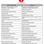
This chart is not intended to be an exhaustive comparison but rather a general overview.

Posted in General.

Tagged with , , , , , , , , , , , , , .


Therapeutic Effects of Bromocriptine

Also see:
Estrogen Increases Serotonin
Anti Serotonin, Pro Libido
Gelatin > Whey
Thyroid peroxidase activity is inhibited by amino acids
Whey, Tryptophan, & Serotonin
Tryptophan, Fatigue, Training, and Performance
Carbohydrate Lowers Free Tryptophan
Protective Glycine
Intestinal Serotonin and Bone Loss
Hypothyroidism and Serotonin
Gelatin, Glycine, and Metabolism
Whey, Tryptophan, & Serotonin
Tryptophan, Sleep, and Depression
Carbohydrate Lowers Serotonin from Exercise
Inflammatory TSH

Antihistamines and some of the antiserotonin drugs (including “dopaminergic” lisuride and bromocriptine) are sometimes useful in cancer treatment, but the safe way to lower serotonin is to reduce the consumption of tryptophan, and to avoid excessive cortisol production (which mobilizes tryptophan from the muscles). Pregnenolone and sucrose tend to prevent over-production of cortisol. -Ray Peat, PhD
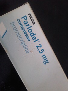
1024141416a

Neuro Endocrinol Lett. 2000;21(5):405-408.
Efficacy of bromocriptine in the treatment of metastatic breast cancer- and prostate cancer-related hyperprolactinemia.
Lissoni P, Mandalà M, Giani L, Malugani F, Secondino S, Zonato S, Rocco F, Gardani G.
OBJECTIVE:
Hyperprolactinemia is a frequent evidence occurring in both metastatic breast cancer and prostate cancer, and it has been proven to be associated with poor prognosis and reduced efficacy of the anticancer therapies. Therefore, the pharmacological control of cancer-related hyperprolactinemia could improve the prognosis of advanced breast and prostate carcinomas. Unfortunately, at present it is still controversial which may be the treatment of cancer-related hyperprolactinemia, which could depend at least in part on a direct autocrine production by cancer cells themselves. The present study was performed to evaluate the acute effects of the long-acting dopaminergic agonist bromocriptine on cancer-related hyperprolactinemia.
METHODS:
The study included 10 women affected by metastatic breast cancer and 10 men with metastatic prostate cancer, showing persistent hyperprolactinemia. Venous blood samples were collected before bromocriptine, and 2, 4, 10 and 24 hours after bromocriptine administration (2.5 mg orally) serum levels of PRL were measured with the double antibody RIA method.
RESULTS:
Bromocriptine induced a normalization of PRL levels in both groups of patients with breast and prostate cancers. Moreover, mean levels of PRL persisted significantly lower than those found before therapy during the whole 24-hour circadian period.
DISCUSSION:
This preliminary study shows that low-dose bromocriptine is sufficient to acutely normalize PRL secretion in both metastatic breast cancer and prostate carcinoma patients, irrespectively of the mechanisms involved in inducing cancer-related hyperprolactinemia. Therefore, low-dose bromocriptine could be recommended in association with the classical antitumor therapies in the treatment of metastatic breast cancer and prostate carcinoma patients showing cancer-related hyperprolactinemia, in an attempt to improve the efficacy of anticancer therapies themselves.

The Lancet, Volume 331, Issue 8586, Pages 609 – 610, 19 March 1988
PERIOPERATIVE BROMOCRIPTINE ADJUVANT TREATMENT FOR OPERABLE BREAST CANCER
I.S. Fentiman , M.A. Chaudary , D.Y. Wang , K. Brame , R.S. Camplhjohn , R.R. Millis
Blood levels of prolactin are consistently raised in women who have undergone mastectomy for breast cancer, probably because of the stress of surgery. Since this increase in concentration of a known breast epithelial growth promoter might stimulate proliferation in micrometastatic cells shed at the time of surgery, a pilot study was conducted to try to abolish this effect. 38 patients with suspected operable breast cancer were given either bromocriptine (18) or placebo (20) tablets for 5 days preoperatively and thereafter for 3-10 days. Bromocriptine-treated patients showed significant reductions in prolactin levels and in S-phase fraction of tumour cells within the primary infiltrating carcinoma. There were no major side-effects. Perioperative bromocriptine may provide another approach to adjuvant therapy, possibly combined with endocrine or cytotoxic treatment for patients with axillary nodal metastases.

The Lancet, Volume 314, Issue 8133, Pages 66 – 69, 14 July 1979
REDUCTION OF PITUITARY-TUMOUR SIZE IN PATIENTS WITH PROLACTINOMAS AND ACROMEGALY TREATED WITH BROMOCRIPTINE WITH OR WITHOUT RADIOTHERAPY
J.A.H. Wass , M.O. Thorner 1, M. Charlesworth , P.J.A. Moult , J.E. Dacie , A.E. Jones , G.M. Besser
69 patients with prolactin-secreting or growth-hormone-secreting pituitary tumours were treated with bromocriptine with or without pituitary irradiation and followed up for 6 months to 6 1/2 years. Of 26 patients with prolactinomas, 11 had external pituitary irradiation in addition to bromocriptine. There was evidence of shrinkage of the pituitary tumour (either a reduction in fossa size or loss of visual-field defects) in 6 of these patients (23%), 3 of whom had been treated with bromocriptine alone. Of 43 acromegalic patients, 30 received external pituitary irradiation. 8 (19%) showed evidence of shrinkage of the pituitary tumour, including 2 who had received no radiotherapy. 1 patient treated with bromocriptine alone showed striking reduction in the size of his suprasellar extension, as assessed by serial computed-tomography scans over 11 months. At the same time his visual-field defects resolved and his deficient corticotrophin and thyrotrophin reserves returned to normal. Bromocriptine can reduce the size of both prolactin-secreting and growth-hormone-secreting pituitary tumours, and this is of potential importance in their management.

The Lancet, Volume 319, Issue 8266, Pages 245 – 249, 30 January 1982
HYPERPROLACTINAEMIA IN MEN—RESPONSE TO BROMOCRIPTINE THERAPY
R.W.G. Prescott a b, P. Kendall-Taylor a b, K. Hall a b, D.G. Johnston a b, A. Crombie a b, A. Mcgregor a b, K. Hall a b
Men with hyperprolactinaemia present with large tumours. Conventional therapy with surgery and/or irradiation is unsatisfactory, with up to 100% of patients remaining hyperprolactinaemic (or subsequently developing pituitary insufficiency). In view of reports of bromocriptine-induced regression of prolactinomas, eight consecutive male hyperprolactinaemic patients with impotence and/or symptoms related to local tumour effects were treated with bromocriptine 20 mg daily as sole therapy for 3-11 months. Symptoms were relieved partly or completely in seven patients and serum prolactin was restored to normal or near normal in all men. Serum thyroxine and plasma cortisol response to hypoglycaemia became normal in two men who had subnormal values before therapy. Mean serum growth hormone response to hypoglycaemia rose significantly as did plasma testosterone concentrations. Evidence of tumour regression, sometimes massive, was seen in the six patients who underwent repeat radiology. The symptomatic relief and biochemical and radiological improvement in these patients indicate that bromocriptine therapy may now be the treatment of choice for hyperprolactinaemic men with large tumours.

The Lancet, Volume 324, Issue 8396, Pages 187 – 192, 28 July 1984
EFFECT OF DOPAMINE AGONIST WITHDRAWAL AFTER LONG-TERM THERAPY IN PROLACTINOMAS
D.G. Johnston , P.Kendall Taylor , M. Watson , K. Hall , D. Patrick , D.B. Cook
The clinical, radiological, and biochemical effects of dopamine agonist withdrawal after long-term treatment were investigated in seven women and eight men who had been treated for prolactinomas for 1·5 to 7 (mean 3·7) years. Before treatment, serum prolactin concentrations were 1473 to 115 000 mU/1, all patients had abnormal radiological findings, and six had suprasellar extensions of pituitary tumours. Treatment with either bromocriptine or pergolide relieved symptoms and suppressed prolactin secretion in most patients. The size of the residual tumour was defined by doing fourth generation computerised . tomographic scans immediately before termination of therapy, and evidence of tumour re-expansion was sought on scans repeated 5-39 weeks later. After discontinuation of treatment, symptoms recurred in 13 of 15 patients and hyper-prolactinaemia redeveloped in 14. Other pituitary function tests remained unchanged or improved. In 13 of 15 patients tumour or gland size did not change after withdrawal of treatment. One man had a marginal increase in tumour size, while in another the pituitary tumour shrank. Thus, although cessation of long-term dopamine agonist therapy leads to recurrence of symptoms and hyperprolactinaemia, rapid tumour regrowth is uncommon and of small extent, and other pituitary function is not altered in the short term.

Br Med J. 1979 September 22; 2(6192): 700–703.
Effects of bromocriptine on pituitary tumour size.
A M McGregor, M F Scanlon, R Hall, and K Hall
In a prospective study designed to assess the influence of bromocriptine on pituitary tumour size 12 patients with pituitary tumours, eight of whom had suprasellar extensions, were treated for three months with 20 mg of bromocriptine daily after a gradual increase to this dose. The group comprised eight women and four men, five with prolactin-secreting adenomas, four with acromegaly, two with functionless adenomas, and one with Nelson’s syndrome. All five patients with prolactin-secreting adenomas showed a reduction in pituitary tumour size as assessed by computerised tomography and metrizamide cisternography accompanied by a fall in prolactin concentrations and clinical and biochemical improvement in their hypopituitarism. One patient in this group had a visual-field defect before treatment, and this resolved. There was no radiological evidence of reduction in tumour size in the remaining seven patients, though this might refect the fairly short duration of treatment, particularly in view of the ancillary evidence of clinical, biochemical, and visual-field improvement in some of the patients. These results emphasise the potential value of bromocriptine in treating patients with large prolactinomas or recurrences of such tumours after previous chiasmal decompression and conventional external megavoltage irradiation on the pituitary.

Br Med J (Clin Res Ed). 1982 June 26; 284(6333): 1908–1911.
Bromocriptine in management of large pituitary tumours.
J A Wass, J Williams, M Charlesworth, D P Kingsley, A M Halliday, I Doniach, L H Rees, W I McDonald, and G M Besser
Bromocriptine has an accepted place in the management of small pituitary tumours that secrete either prolactin or growth hormone. The treatment of large tumours with extrasellar extensions is more difficult, however: though surgery is the standard treatment, it is often unsuccessful in returning excessive hormone secretion to normal and may cause hypopituitarism. A prospective trial was undertaken to assess the frequency with which changes in pituitary function and size of large tumours occurs. Nineteen patients were studied before and during treatment with bromocriptine (7.5 to 60 ml/day) for three to 22 months, using contrast radiology and a detailed assessment of pituitary function. Eighteen patients had hyperprolactinaemia and two of these also had raised concentrations of growth hormones; one patient had an apparently non-functioning tumour. In 12 patients (63%) tumour size decreased with bromocriptine and no tumour enlarged. Nine patients had visual-field defects, which improved in seven, becoming normal in five. Pituitary function improved in nine patients (47%) becoming entirely normal in three. Bromocriptine should be the treatment of choice in patients with large pituitary tumours with extrasellar extensions, provided close supervision is maintained.

JAMA. 1982 Jan 15;247(3):311-6.
Bromocriptine reduces pituitary tumor size and hypersection. Requiem for pituitary surgery?
Spark RF, Baker R, Bienfang DC, Bergland R.
Twelve patients with pituitary tumor whose prior treatment included surgery and radiotherapy in four, surgery alone in four, radiotherapy alone in one, and none in three were studied. Nine had hyperprolactinemia, two had elevated serum growth hormones, and one had no pituitary hormone excess. Visual field defects were present in six. All had pituitary-gonadal insufficiency manifested as impotence or amenorrhea. All were tested with bromocriptine, 7.5 to 25 mg daily, and followed up for eight to 27 (mean 15) months. Serum prolactin levels decreased to normal in seven of nine patients. Serum growth hormone values were normalized in both acromegalics. When hormone levels were reduced to normal, pituitary tumor size decreased. Vision was restored to normal in five of six patients, including one patient with pituitary tumor but no pituitary hormone excess. Bromocriptine corrects the physiological defects associated with pituitary tumors that have been incompletely treated with surgery, radiotherapy, or both and may be a useful primary treatment for patients with pituitary tumors.

Am J Med. 1983 Nov;75(5):868-74.
Hyperprolactinemia. Long-term effects of bromocriptine.
Johnston DG, Prescott RW, Kendall-Taylor P, Hall K, Crombie AL, Hall R, McGregor A, Watson MJ, Cook DB.
Patients with hyperprolactinemia may be managed by pituitary surgery or irradiation, bromocriptine treatment, or a combination of these methods, and some patients remain untreated. Little is known of the long-term consequences of some of these therapeutic regimens. Forty-six hyperprolactinemic patients (40 female and six male) managed solely with bromocriptine or no treatment over a period of 12 months to six years were therefore evaluated in this study. Nine patients with radiologically normal pituitary fossae were untreated and 10 received bromocriptine, 7.5 to 10 mg daily, while 20 patients with radiologic evidence of a pituitary tumor were treated with bromocriptine, generally 10 to 20 mg daily. Patients were assessed clinically, biochemically, and radiologically before treatment and at least six weeks after discontinuation of therapy. A further seven patients were similarly assessed before and after eight bromocriptine-induced pregnancies. Symptoms persisted in the untreated group of nine patients, although menstruation returned in four of the females with previous amenorrhea; serum prolactin levels remained elevated, other pituitary function did not change, and pituitary fossae remained normal radiologically. In all patients treated with bromocriptine, symptoms improved irrespective of radiologic findings on the pituitary, and were abolished in 67 percent during treatment associated with a decrease in serum prolactin levels in all, and a return of levels to within normal limits in 80 percent of patients. Persistent side effects were usually dose-related, but remained troublesome in 13 percent. Bromocriptine-induced tumor regression was evident radiologically in all patients with suprasellar tumor tissue and in some with purely intrasellar adenomas. This effect occurred rapidly and persisted or increased throughout follow-up. On discontinuation of treatment, prolactin levels remained significantly lower than before therapy (mean 2,934 versus 5,052 mU/liter, p less than 0.05) but were within the normal range in only two patients. Other pituitary function was unaltered, or improved in some patients with definite tumors. Bromocriptine-induced pregnancy produced no permanent change in clinical, biochemical, or radiologic status. Long-term bromocriptine treatment for hyperprolactinemia is thus highly effective in alleviating symptoms and suppressing prolactin secretion, and induces persistent tumor regression on treatment without deterioration of other pituitary function in patients with macroadenomas. On discontinuation of therapy, however, hyperprolactinemia usually recurs, and treatment may therefore need to be continued for years.

Am J Dis Child. 1993 Oct;147(10):1057-61.
Prolactin-secreting macroadenomas in adolescents. Response to bromocriptine therapy.
Tyson D, Reggiardo D, Sklar C, David R.
OBJECTIVE:
To report five cases of prolactin (PRL)-secreting macroadenomas in adolescents, including their presentations and responses to bromocriptine mesylate treatment.
PATIENTS:
Five adolescents (three females and two males) aged between 12.5 and 17 years were diagnosed as having PRL-secreting macroadenomas at the pediatric endocrine service at New York University Medical Center between 1987 and 1989. Presenting complaints included visual field deficits, gynecomastia, and amenorrhea, both primary and secondary. All patients demonstrated some feature of hypogonadism or pubertal arrest. Diagnostic criteria included an elevated serum PRL level (mean, 1670 micrograms/L; range, 610 to 3700 micrograms/L) and visualization of a pituitary tumor that measured greater than 1 cm by either a computed tomographic scan or magnetic resonance imaging (mean size, 2.7 cm; range, 1.4 to 4 cm).
INTERVENTIONS:
Each patient was treated with bromocriptine mesylate at an oral dose of 7.5 mg/d. The patients continued with that treatment for the duration of the study period.
MEASUREMENTS AND RESULTS:
Anterior pituitary function was evaluated in four of five patients before treatment. All four were growth hormone deficient. Three patients were also gonadotropin deficient. Thyrotropin (thyroid-stimulating hormone) and corticotropin (adrenocorticotropic hormone) deficiencies were demonstrated in three patients who had multiple pituitary deficits. Follow-up testing included serial PRL measurements and radiographic imaging of tumor size. All patients demonstrated a marked decrease in PRL levels, as well as in tumor size (mean shrinkage, 70%). The three patients who initially had visual field deficits showed significant improvement of vision with bromocriptine therapy. Follow-up study of anterior pituitary function showed significant improvement with bromocriptine treatment in three patients.
CONCLUSIONS:
Bromocriptine was quite effective in the shrinkage of PRL-secreting macroadenomas in all our patients. It is a noninvasive treatment that can preserve and restore vision, as well as pituitary function, which is integral to continued growth and sexual maturation of the adolescent. Bromocriptine is preferable to surgery or radiation in the treatment of PRL-secreting macroadenomas in the adolescent.

Ups J Med Sci. 1982;87(3):259-67.
Rapid regression of pituitary tumours during bromocriptine treatment of women with hyperprolactinaemia.
Bergh T, Nillius SJ, Lundberg PO, Moström U, Enoksson P, Ohman L, Wide L.
Four hyperprolactinaemic women with large pituitary adenomas with suprasellar extension were given primary tumour therapy with bromocriptine. The treatment resulted in rapid tumour regression in all the women, as verified by repeated computerized tomography (CT) scans. Pronounced visual field defects were present in three of the four women before treatment. All of them had marked improvement of vision within a few days after the initiation of bromocriptine therapy and they regained normal or nearly normal visual fields during the treatment. The raised serum prolactin concentrations decreased to normal levels in all the women. Thus, medical treatment with bromocriptine can induce rapid tumour regression in patients with hyperprolactinaemia and large pituitary tumours.

Obstet Gynecol. 1989 Mar;73(3 Pt 2):517-20.
Successful treatment of a prolactin-producing pituitary macroadenoma with intravaginal bromocriptine mesylate: a novel approach to intolerance of oral therapy.
Katz E, Schran HF, Adashi EY.
A 37-year-old woman with a symptomatic 18-mm prolactin-secreting pituitary adenoma could not be managed with oral bromocriptine mesylate because of unacceptable gastrointestinal side effects. However, when given the medication intravaginally, the patient was successfully treated as assessed by evident tumor shrinkage and diminished secretory activity. Serum bromocriptine levels were approximately six to eight times higher than those reported after oral administration. The vaginal route may help reduce some of the adverse effects of bromocriptine mesylate experienced during oral administration and may possibly allow lowering of the overall effective dose by avoiding first liver passage.

Research on LSD and its derivatives led to drugs such as bromocriptine, which oppose the effects of histamine and estrogen. Some of bromocriptine’s effects are clearly antagonistic to serotonin, though bromocriptine is usually called a “dopamine agonist”; dopamine is pretty generally a serotonin antagonist. -Ray Peat, PhD

Eur J Pharmacol. 1982 Jul 30;81(4):569-76.
Actions of serotonin antagonists on dog coronary artery.
Brazenor RM, Angus JA.
Serotonin released from platelets may initiate coronary vasospasm in patients with variant angina. If this hypothesis is correct, serotonin antagonists without constrictor activity may be useful in this form of angina. We have investigated drugs classified as serotonin antagonists on dog circumflex coronary artery ring segments in vitro. Ergotamine, dihydroergotamine, bromocriptine, lisuride, ergometrine, ketanserin, trazodone, cyproheptadine and pizotifen caused non-competitive antagonism of serotonin concentration-response curves. In addition, ketanserin, trazodone, bromocriptine and pizotifen inhibited noradrenaline responses in concentrations similar to those required for serotonin antagonism. All drugs with the exception of ketanserin, cyproheptadine and pizotifen showed some degree of intrinsic constrictor activity. Methysergide antagonized responses to serotonin competitively but also constricted the coronary artery. The lack of a silent competitive serotonin antagonist precludes a definite characterization of coronary serotonin receptors at this time. However, the profile of activity observed for the antagonist drugs in the coronary artery differs from that seen in other vascular tissues. Of the drugs tested, ketanserin may be the most useful in variant angina since it is a potent 5HT antagonist, lacks agonist activity and has alpha-adrenoceptor blocking activity.

In an animal study, bromocriptine, which shifts the balance away from serotonin, reduced obesity and insulin and free fatty acids, and improved glucose tolerance. -Ray Peat, PhD

Neuroendocrinology. 1998 Jul;68(1):1-10.
Bromocriptine reduces obesity, glucose intolerance and extracellular monoamine metabolite levels in the ventromedial hypothalamus of Syrian hamsters.
Luo S, Meier AH, Cincotta AH.
We examined whether reductions in body fat stores and insulin resistance in Syrian hamsters induced by bromocriptine are associated with reductions in daily norepinephrine (NE) and serotonin activities as indicated by their extracellular metabolite levels in the ventromedial hypothalamus (VMH). High levels of these monoamines within the VMH have been suspected to induce obesity and insulin resistance. Microdialysate samples from the VMH of freely moving obese male hamsters (BW: 208 +/- 5 g) were collected hourly over a 25-hour period before bromocriptine treatment, during the first day of and after 2 weeks of bromocriptine treatment (800 microg/animal daily, i.p.), and body composition and glucose tolerance analyses were conducted before and after 2 weeks of treatments. The microdialysate samples were analyzed by HPLC for metabolites of serotonin: 5-hydroxy-indoleacetic acid (5-HIAA), NE: 3-methoxy-4-hydroxy-phenylglycol (MHPG), and dopamine: homovanillic acid (HVA). Bromocriptine treatment for 14 days significantly reduced body fat by 60% and areas under the glucose and insulin curves during a glucose tolerance test by 50 and 46%, respectively. Concurrently, extracellular VMH contents of 5-HIAA, MHPG, and HVA were reduced by 50, 29 and 66%, respectively (p < 0.05). Similarly, VMH 5-HIAA and MHPG contents were 48 and 44% less, respectively (p < 0.05), in naturally glucose-tolerant hamsters compared with naturally glucose-intolerant hamsters. Bromocriptine induced reductions of body fat, and improvements in glucose intolerance may result in part from its ability to decrease serotonin and NE activities in the VMH.

Metabolism. 1991 Jun;40(6):639-44.
Bromocriptine inhibits the seasonally occurring obesity, hyperinsulinemia, insulin resistance, and impaired glucose tolerance in the Syrian hamster, Mesocricetus auratus.
Cincotta AH, Schiller BC, Meier AH.
Seasonally obese-hyperinsulinemic female Syrian hamsters were injected daily with bromocriptine or saline for a period of 34 days to test for effects of bromocriptine on body fat store levels, hepatic triglyceride secretion, glucose tolerance, and plasma insulin and glucose concentrations. The effects of bromocriptine on body fat store levels, as well as on plasma insulin and glucose concentrations, in seasonally obese hamsters were compared with the levels of body fat, plasma insulin, and plasma glucose observed in seasonally lean hamsters. Bromocriptine treatment substantially improved glucose intolerance and reduced the total and stimulated areas under the glucose tolerance curve by 33% after 14 days of treatment. After 34 days of treatment, bromocriptine reduced body fat store levels by 36% and hepatic triglyceride secretion by 40% without any concurrent change in food consumption. Furthermore, bromocriptine reduced the plasma insulin level by 70%, while slightly reducing plasma glucose concentration (ie, 68% reduction in the insulin to glucose ratio). The reductions of body fat, plasma insulin, and plasma insulin to glucose ratio produced by bromocriptine in seasonally obese hamsters are equivalent to those observed in seasonally lean hamsters. Shifts in phase relationships of circadian neuroendocrine rhythms have been demonstrated to regulate annual cycles of metabolism in vertebrates, including the Syrian hamster. The effects of bromocriptine can also be explained as an alteration of such a circadian mechanism.

Metabolism. 1995 Oct;44(10):1349-55.
Bromocriptine inhibits in vivo free fatty acid oxidation and hepatic glucose output in seasonally obese hamsters (Mesocricetus auratus).
Cincotta AH, Meier AH.
Seasonally obese hyperinsulinemic hamsters were treated for 5 weeks with bromocriptine (500 to 600 micrograms per animal) and tested for drug effects on energy balance, body fat stores, nocturnal whole-body free fatty acid (FFA) metabolism and hepatic glucose output, and diurnal glucose tolerance. After 5 weeks, bromocriptine treatment reduced retroperitoneal fat pad weight by 45% without altering either daily food consumption or end-treatment total daily energy expenditure. Also, 5 weeks of treatment improved the diurnal glucose tolerance, resulting in a 47% and 33% decrease in the area under glucose and insulin curves, respectively. After 4 weeks, bromocriptine treatment reduced nocturnal lipolysis by 28%, palmitate rate of appearance into plasma by 30%, palmitate oxidation by 33%, and hepatic glucose output by 28%. Moreover, these reductions were accompanied by a 75% reduction in plasma insulin concentration. The data suggest that bromocriptine may improve diurnal glucose tolerance in part by inhibiting the preceding nocturnal lipolysis and FFA oxidation. Reductions in nocturnal FFA oxidation and hepatic glucose production may result from bromocriptine’s influences on circadian organization of hypothalamic centers known to regulate these activities. Available evidence suggests that bromocriptine may impact this neuroendocrine organization of metabolism by increasing the dopamine to noradrenaline activity ratio in central (hypothalamic) and peripheral (eg, liver and adipose) target tissues.

DIABETES CARE, VOLUME 20, NUMBER 11, NOVEMBER 1997
Effects of a Quick-Release Form of Bromocriptine (Ergoset) on Fasting and Postprandial Plasma Glucose, Insulin, Lipid, and Lipoprotein Concentrations in Obese Nondiabetic Hyperinsulinemic Women
Kamath and Associates
OBJECTIVE — To assess the effect on various aspects of carbohydrate and lipid metabolism of administering a quick-release formulation of bromocriptine (Ergoset) to obese, nondiabetic, hyperinsulinemic women.
RESEARCH DESIGN AND METHODS— Hourly concentrations of prolactin, glucose, insulin, free fatty acid (FFA), and triglyceride were measured for 24 h before and after approximately 8 weeks of treatment with Ergoset. In addition, fasting lipid and lipoprotein concentrations and the steady-state plasma glucose (SSPG) concentration in response to a continuous infusion of somatostatin, insulin, and glucose were determined before and after Ergoset administration.
RESULTS — Circulating prolactin concentrations were dramatically decreased (P < 0.001) following treatment, associated with a significant fall (P < 0.05) in 24-h-long plasma glucose, FFA, and triglyceride concentrations. Neither circulating plasma insulin concentrations nor the ability of insulin to mediate glucose disposal changed with treatment. Finally, fasting total cholesterol fell (P < 0.05) and the ratio of total to HDL cholesterol decreased (P = 0.06) in association with Ergoset treatment. CONCLUSIONS — The fact that significant metabolic improvement was seen in the obese nondiabetic hyperinsulinemic women studied suggests that Ergoset could be of therapeutic benefit in clinical conditions of hyperglycemia and/or dyslipidemia.

J Hepatol. 2006 Sep;45(3):439-44. Epub 2006 May 6.
Bromocriptine reduces steatosis in obese rodent models.
Davis LM, Pei Z, Trush MA, Cheskin LJ, Contoreggi C, McCullough K, Watkins PA, Moran TH.
BACKGROUND/AIMS:
Obesity is a risk factor for glucose intolerance, steatosis, and oxidative stress, characteristics of nonalcoholic fatty liver disease. Bromocriptine may have anti-obesity, insulin-sensitizing, lipolytic, and antioxidant properties. We, therefore, hypothesized that bromocriptine would improve markers of nonalcoholic fatty liver disease in obese rodent models.
METHODS:
We performed a randomized, controlled experiment in genetically obese fatty Zucker rats and diet-induced obese rats to assess for behavioral and peripheral anti-obesity actions of bromocriptine (10mg/kg) that would improve nonalcoholic fatty liver disease.
RESULTS:
Behaviorally, food intake decreased and locomotor activity increased in bromocriptine-treated fatty Zucker and dietary-induced obese rats. Peripherally, liver triglycerides were significantly reduced and hepatic manganese superoxide dismutase significantly increased in bromocriptine-treated fatty Zucker and diet-induced obese rats compared to controls. Blood glucose was significantly lower in bromocriptine-treated Zucker rats compared to fatty controls and was no different than that of lean controls.
CONCLUSIONS:
Improvements in obesigenic behaviors, glucose tolerance, hepatic lipid accumulation, and mitochondrial oxidative stress observed in genetically obese and diet-induced obese rodents indicate that bromocriptine may be promising as a broad-based therapy for nonalcoholic fatty liver disease.

Eur J Pharmacol. 1984 Mar 16;99(1):85-90.
One minute of bromocriptine irreversibly inhibits prolactin release for hours.
Cronin MJ, Evans WS, Thorner MO.
The ability of the dopamine receptor antagonist spiperone to block dopamine- or bromocriptine-inhibited prolactin release from dispersed rat anterior pituitary cells was tested in vitro. In a continuously perfused cell column apparatus, spiperone rapidly counteracted the inhibitory effect of dopamine but was unable to reverse the inhibitory effect of the potent dopamine agonist bromocriptine when added 30 min after bromocriptine. However, spiperone completely blocked the bromocriptine action if added simultaneously with bromocriptine. These basic data were confirmed, and the time relationships more accurately defined, in static incubations of monolayer cultures. Spiperone blocked the typical inhibition of prolactin release by bromocriptine only if the cells were pre- or coincubated with the antagonist. If spiperone was added as soon as 1 min after bromocriptine, the antagonist was unable to block the complete expression of the bromocriptine inhibition. These results suggest that bromocriptine is a functionally irreversible dopamine agonist for at least the 4 h of these studies.

Proc Soc Exp Biol Med. 1984 Feb;175(2):191-5.
Bromocriptine inhibits growth hormone release from rat pituitary cells in primary culture.
Cronin MJ, Thorner MO, Hellmann P, Rogol AD.
The action of the potent dopamine receptor agonist bromocriptine was studied in primary cultures of rat anterior pituitary cells. Bromocriptine inhibited both prolactin and growth hormone release in a concentration-dependent manner. This effect was blocked by the dopamine receptor antagonist spiperone when the agonist and antagonist were added coincidently. In contrast, spiperone was unable to affect the actions of bromocriptine if added 1 min after bromocriptine application or later. These results suggest that dopamine receptors exist not only on mammotrophs, but also on somatotrophs in vitro.

J Neural Transm. 1981;51(1-2):61-82.
The endocrine profile of bromocriptine: its application in endocrine diseases.
Lancranjan I.
Bromocriptine, a potent agonist at Dz receptors, was developed as a therapeutic agent for inhibiting prolactin (PRL) secretion in patients with hyperprolactinemia. Besides, its PRL-suppressive effect and a short-lasting growth hormone (GH)-releasing effect in normal volunteers, bromocriptine has no other endocrine effects in healthy subjects. On the other hand, bromocriptine lowers GH secretion in acromegalic patients and ACTH secretion in some patients with Cushing’s disease or Nelson’s syndrome. The paper reviews the endocrine actions of bromocriptine in man, in normal and pathological conditions, the bromocriptine’s mechanism of action and its clinical applications in endocrinology.

Diabetes Care. 1996 Jun;19(6):667-70.
Bromocriptine (Ergoset) reduces body weight and improves glucose tolerance in obese subjects.
Cincotta AH, Meier AH.
OBJECTIVE:
A double-blind placebo controlled study investigated long-term effects of Ergoset, a new quick release formulation of bromocriptine, on body weight, body fat, and glucose tolerance in a group (n = 17) of obese subjects who were instructed to follow a moderate hypocaloric diet.
RESEARCH DESIGN AND METHODS:
Obese individuals (> 25% body fat for men and > 30% body fat for women) were instructed to follow a calorie-restricted diet (70% of weight maintaining based on study entry weight) and were randomized to daily treatment with Ergoset (1.6-2.4 mg/day) or placebo at 0800 over an 18-week treatment period. Oral glucose tolerance tests were performed on subjects before initiation and again at termination of treatment. Body weight and body fat (determined by skinfold measurements) were quantified every 2 weeks during the course of treatment.
RESULTS:
Ergoset treatment for 18 weeks significantly reduced body weight and body fat versus placebo (6.3 +/- 1.5 and 5.4 +/- 1.1 kg vs. 0.9 +/- 1.0 and 1.5 +/- 0.6 kg. respectively, P < 0.01). Ergoset, but not placebo, also improved glucose tolerance (P < 0.02); the stimulated area under the oral glucose tolerance curve was reduced by 46% (from 121 +/- 23 to 64 +/- 32 mg.h-1.dl-1), while the stimulated area under the insulin curve was reduced by 30%.
CONCLUSIONS:
When combined with instruction to follow a moderate hypocaloric diet, Ergoset, but not placebo, improves glucose tolerance and promotes significant weight and body fat loss in obese subjects over an 18- week treatment period.

Diabetes Care. 2000 Aug;23(8):1154-61.
Bromocriptine: a novel approach to the treatment of type 2 diabetes.
Pijl H, Ohashi S, Matsuda M, Miyazaki Y, Mahankali A, Kumar V, Pipek R, Iozzo P, Lancaster JL, Cincotta AH, DeFronzo RA.
OBJECTIVE:
In vertebrates, body fat stores and insulin action are controlled by the temporal interaction of circadian neuroendocrine oscillations. Bromocriptine modulates neurotransmitter action in the brain and has been shown to improve glucose tolerance and insulin resistance in animal models of obesity and diabetes. We studied the effect of a quick-release bromocriptine formulation on glucose homeostasis and insulin sensitivity in obese type 2 diabetic subjects.
RESEARCH DESIGN AND METHODS:
There were 22 obese subjects with type 2 diabetes randomized to receive a quick-release formulation of bromocriptine (n = 15) or placebo (n = 7) in a 16-week double-blind study. Subjects were prescribed a weight-maintaining diet to exclude any effect of changes in body weight on the primary outcome measurements. Fasting plasma glucose concentration and HbA(1c) were measured at 2- to 4-week intervals during treatment. Body composition (underwater weighing), body fat distribution (magnetic resonance imaging), oral glucose tolerance (oral glucose tolerance test [OGTT]), insulin-mediated glucose disposal, and endogenous glucose production (2-step euglycemic insulin clamp, 40 and 160 mU x min(-1) x m(-2)) were measured before and after treatment.
RESULTS:
No changes in body weight or body composition occurred during the study in either placebo- or bromocriptine-treated subjects. Bromocriptine significantly reduced HbA(1c) (from 8.7 to 8.1%, P = 0.009) and fasting plasma glucose (from 190 to 172 mg/dl, P = 0.02) levels, whereas these variables increased during placebo treatment (from 8.5 to 9.1%, NS, and from 187 to 223 mg/dl, P = 0.02, respectively). The differences in HbA(1c) (delta = 1.2%, P = 0.01) and fasting glucose (delta = 54 mg/dl, P < 0.001) levels between the bromocriptine and placebo group at 16 weeks were highly significant. The mean plasma glucose concentration during OGTT was significantly reduced by bromocriptine (from 294 to 272 mg/dl, P = 0.005), whereas it increased in the placebo group. No change in glucose disposal occurred during the first step of the insulin clamp in either the bromocriptine- or placebo-treated group. During the second insulin clamp step, bromocriptine improved total glucose disposal from 6.8 to 8.4 mg x min(-1) kg(-1) fat-free mass (FFM) (P = 0.01) and nonoxidative glucose disposal from 3.3 to 4.3 mg min(-1) x kg(-1) FFM (P < 0.05), whereas both of these variables deteriorated significantly (P < or = 0.02) in the placebo group.
CONCLUSIONS:
Bromocriptine improves glycemic control and glucose tolerance in obese type 2 diabetic patients. Both reductions in fasting and postprandial plasma glucose levels appear to contribute to the improvement in glucose tolerance. The bromocriptine-induced improvement in glycemic control is associated with enhanced maximally stimulated insulin-mediated glucose disposal.

Acta Neurol Scand. 1977 Jul;56(1):37-45.
Treatment of Huntington’s chorea with bromocriptine.
Frattola L, Albiazzati MG, Spano PF, Trabucchi M.
The authors tested the effects of 2-Br-ergocriptine (bromocriptine, CB-154), a drug which exerts a mixed agonist-antagonist activity on the dopaminergic receptors, in 12 patients with Huntington Chorea in a double-blind crossover trial. This treatment significantly reduced the abnormal involuntary movements and the disease severity in most of the patients. Subjects who were slightly disabled showed a better response than the ones with more severe degrees of disability.

Obstet Gynecol. 1980 Mar;55(3):278-84.
Bromocriptine mesylate (Parlodel) in the management of amenorrhea/galactorrhea associated with hyperprolactinemia.
Cuellar FG.
The efficacy and safety of bromocriptine mesylate (5 to 7.5 mg per day for up to 24 weeks) were studied in 22 clinical trials involving 226 patients who had amenorrhea/galactorrhea associated with hyperprolactinemia and no demonstrable pituitary tumor. Of the 187 patients evaluated for efficacy, 80% experienced reinitiation of menses (or pregnancy without first having menses); the average treatment time (excluding those who conceived) was 5.7 weeks. Galactorrhea was significantly (at least 75%) reduced in 76% of the 187 patients after an average treatment tome of 6.4 weeks, and was completely suppressed in 58% after 12.7 weeks. Maximum reduction in serum prolactin levels occurred within the first 4 weeks of therapy and the reduced levels were maintained during treatment; moreover, there was a strong correlation between prolactin reduction and clinical improvement. Adverse reactions were reported by 68% of the 226 patients evaluated for safety; in general, these reactions were mild and transient. Several patients experienced hypotension, but only 1 discontinued therapy because of it. Based on these findings, bromocriptine mesylate was judged safe and efficacious for this purpose.

Posted in General.

Tagged with , , , , , , , , , , , , , , .


Burzynski: Cancer Is Serious Business

Posted in General.

Tagged with , .