Categories:

Bottled Water 2011: Summary of Findings

Study authors: Nneka Leiba, MPH, Analyst; Sean Gray, MS, Senior Analyst; Jane Houlihan, MSCE, Sr VP for Research

Overview

The bottled water industry routinely fails to provide information to consumers about the water’s specific geographic source, purification methods and the results of purity testing, a new EWG investigation shows.

Overall, more than half of the 173 bottled water brands surveyed in 2010 flunked EWG’s transparency test.

Many brands fill their labels with vague claims of a pristine source or perfect purity — but no real facts. If people are willing to pay up 1,900 times the cost of tap water in order to buy water in a plastic bottle, they deserve better than that1.

EWG’s last label survey (2009) found that only two of 188 bottled water brands provided the three most basic facts about their water — source name and location, treatment and purity. Since then, the Government Accountability Office has taken the industry and the federal Food and Drug Administration to task for lax inspection and disclosure practices. During a heavily publicized Congressional hearing on the GAO and EWG reports, House subcommittee chairman Bart Stupak, D-Mich., declared, “Just because it comes in a bottle, we assume it’s healthier, but it’s not the case.”

EWG’s 2010 survey shows that 18 percent of bottled water brands still fail to reveal their water’s geographic source; 32 percent are mum on treatment methods and purity testing; and 13 percent publish “water quality” reports that lack any actual testing results.

More than half of the brands EWG surveyed either made no improvements in transparency — or revealed even less in 2010 than in 2009. Bottled water sales continue to be fueled by expensive marketing and misinformation, while many companies stubbornly hide the truth about what’s in the bottle.

When it comes to transparency about bottled water, here are the best and the worst:

  • Best — From 2009 to 2010, the number of brands getting top grades for transparency rose from just two all the way to… three — hardly better. These three – Gerber Pure Purified Water, Nestle Pure Life Purified Water, and Penta Ultra-Purified Water – earned the highest possible marks for labeling the location of their water source and treatment methods and posting online reports on purity. Even so, the water quality reports for all three are outdated, with lab tests dating to 2008.
  • Worst — Six brands, including Whole Foods’ Italian Still Mineral Water, sank to the bottom of the barrel. They provide consumers with none of the three basic facts about their water, either on labels or on company websites.

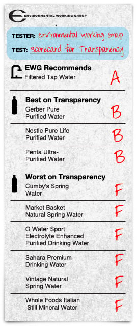
Each “best on transparency” bottled water brand shows a specific geographic location of its water source and treatment method on the label and posts purity testing online. The “worst on transparency” bottled waters list no information on the water’s source location, treatment or purity, online or on the label. These lists are drawn from EWG’s survey of labels from 173 bottled waters purchased in 2010.

America’s bottled water habit has consequences: every 27 hours Americans drink enough bottles of water to circle the equator with empty plastic containers.2

Bottled water companies enjoying this massive commercial success may suspect that their customers would turn away if they knew that most of them draw their product from municipal tap water (BMC 2010, Food and Water Watch 2010), or that the plastics used to make the bottles can be laced with chemical additives that leach into the water (EWG 2008). Perhaps that’s why EWG’s survey found:

  • Eleven companies disclosed less in 2010 than in 2009. Crystal Geyser, Sam’s Choice and nine other companies or brands disclosed even less information in 2010 than in 2009, for one or more bottled waters they sell.[See all products disclosing less in 2010 than in 2009]
  • Twenty-nine brands have ignored California’s new disclosure law. More than a quarter of the water bottles purchased in California did not list certain consumer information on the label or failed to provide a water quality report when contacted by EWG as are required under state law. These brands include Fiji Natural Artesian Water and Green Planet Pure Handcrafted Water.[read more]
  • Eight of the 10 top-selling domestic brands earned a D or F for transparency. Another earned an unimpressive C. These nine brands don’t label the specific location of their water source and treatment method or provide contact information for consumers to get information on water purity. They include Pepsi’s Aquafina brand, Coca-Cola’s Dasani, Crystal Geyser and six of seven brands produced by Nestlé Waters NA. Of the 10 top domestic brands, only Nestlé’s Pure Life Purified Water brand lists a specific water source and treatment method on the label and provides a water quality testing report upon request.

Three basic facts – source, treatment, purity – remain hidden

EWG supports efforts by states such as California, Massachusetts, and New Mexico to supplement federal law when it comes to requiring companies to disclose more consumer information about their bottled water products. The fact is, however, that secrecy remains common and is perfectly legal in many states. Among the bottled water labels surveyed by EWG:

18 percent did not list the water’s geographic source on label or website.

EWG’s 2010 survey found that many companies choose not to disclose the location of their water source. Of 173 brands surveyed, 32 (18 percent), including Publix, Kroger and Harris Teeter store brands, do not provide this information on either their label or their website.[see all 32]

Of brands included in both EWG’s 2009 and 2010 surveys, 18 did not label their water source location in either 2009 or 2010. They include Dasani Purified Water, Glaceau’s Smartwater, Kroger Purified Drinking Water and 15 others.[see all 18]

32 percent provide no information on water treatment.

Producers of 55 brands (32 percent), including Giant’s Acadia Natural Spring Water and CVS’ Gold Emblem Natural Spring Water, gave consumers no way to learn the purity of their water. These companies fail to disclose any information about their treatment methods or do not post a water quality report online. [see all 55]

Of brands surveyed by EWG in both 2009 and 2010, 30 failed to provide this information both years. They included Fiji, Evian, Trader Joe’s and Nestlé Waters. Their customers may be swallowing municipal water bottled straight from the tap – or pumped from a well to a truck to a bottle – and not purified at all. [see all 30]

13 percent provide “water quality” reports lacking actual test data.

Twenty-two bottled waters (13 percent) publish water quality reports that contain no testing results, including Safeway’s Refreshe and Walmart’s Great Value brands, among many others. [see all 22]

Is the industry telling you more in 2010? Barely.

How many brands offered more information on source, treatment, and purity?
(2010 vs. 2009)

Source: Environmental Working Group analysis of bottled water labels and websites and personal communication with companies. Categories defined as follows — Better: labels surveyed were more transparent in 2010 than in 2009. Mixed: some new information was listed, but other data provided in 2009 were now missing. No progress: no change in transparency since 2009. Worse: some or all labels surveyed were less transparent than in 2009.

EWG compared labels from 2009 and 2010 for 72 brands to see whether companies were providing more basic information to their customers. We expected progress. After all, listing the site of their water source and treatment method on the label is a simple job. Posting a water quality report on a company website also presents few hurdles. And following last year’s Congressional hearing, GAO report and tremendous pressure from consumer groups, you might expect even the most resistant companies to budge, at least a little.

The results are disappointing. Some brands disclosed even less information in 2010 than in 2009, and only a fraction were more open.

Overall, bottled water companies stack up as follows:

Getting worse

11 brands (15 percent) disclosed less in 2010 than in 2009 (click for “Getting worse” and “Some decline” lists).

Eleven companies disclosed less about their water in 2010 than in 2009, including Nursery Purified Water and Sam’s Choice Purified Drinking Water.

No progress

27 brands (38 percent) disclosed no more in 2010 than in 2009.

These brands might easily have listed the location of their water source, treatment methods and testing data, but instead continue to hide much of this information, just as they did last year.

Better

28 brands (39 percent) disclosed more in 2010 than in 2009 on some or all labels surveyed for each product (click for “Better” and “Some improvement” lists).

Brands providing more information about the specific geographic location of their source, treatment and purity in 2010 than in 2009 included Acqua Panna and Mountain Valley. Last year Nestlé Pure Life’s label listed multiple possible water source locations; the 2010 labels named the specific location of the source. Vasa and San Pellegrino added information on labels for consumers wishing to get additional information on the water’s quality.

Major brands obscure basic data about their products

Large and small brands alike withhold basic information about their products. Labels of nine of the 10 top-selling domestic brands do not identify their specific water source or treatment method or provide contact information for consumers seeking additional information on water quality. These big brands include Pepsi’s Aquafina, Coca-Cola’s Dasani, Crystal Geyser and six of seven brands produced by Nestlé Waters NA. Of the 10 top domestic brands, only Nestlé’s Pure Life Purified Water lists a specific water source and treatment method on the label and provides a water quality testing report upon request. Here are some of the grades major brands earned on EWG’s scorecard:

  • Pepsi’s Aquafina Purified Drinking Water The label says the water “originates from public water sources” but fails to name them. The water is treated with a process called “HydRO-7™” that is not explained on the label. Only three of the 10 Aquafina labels assessed list a phone number for consumers seeking more information on water quality. Even with the phone number, obtaining a water quality report may not be possible; a company representative told EWG that water quality testing information was “proprietary.”
  • Nestlé’s Arrowhead Mountain Spring Water lists a number of California springs as possible sources for the products EWG assessed. The labels do not include any information on how the water is treated but do list a phone number and website for consumers seeking water quality information.
  • CG Roxane’s Crystal Geyser Natural Alpine Spring Water lists a number of “CG Roxane Source[s]” for the water EWG obtained but offered no specific names of springs. The labels provide no information on treatment, and a third of them do not direct consumers how to get more information on water quality.
  • Coca-Cola’s Dasani Purified Water does not name its source’s geographic site on the label, but notes that the water is treated by reverse osmosis. Six of the seven labels surveyed direct consumers to additional water quality information.
  • Nestlé’s Deer Park Natural Spring Water lists a number of springs in Pennsylvania, Florida, Maine, Tennessee and Maryland as possible water sources on the labels EWG assessed. No treatment method is listed and none of the labels give consumers a contact to get information on water quality.
  • Nestlé’s Ice Mountain Natural Spring Water’s label lists two springs in Michigan as possible sources but fails to describe its treatment methods. None of the labels give consumers a contact to get information on water quality.
  • Nestlé Pure Life Purified Water’s label indicates that the source is either “deep protected wells” in Florida, Michigan or California or the public water supplies of specified cities in Pennsylvania, Colorado or Florida. The water is treated either by reverse osmosis or distillation, and all the labels include contact information for consumers seeking additional information on water quality.
  • Nestlé’s Ozarka Natural Spring Water label says the water is “a blend of Roher Spring, Henderson County, TX and Piney Wood Springs, Wood County, TX” but it does not list treatment methods or include contact information for requesting water quality reports.
  • Nestlé’s Poland Spring Natural Spring Water’s label lists a number of springs in Maine as possible sources but does not identify treatment methods or contacts for water quality information.
  • Nestlé’s Zephyrhills Natural Spring Water’s label lists four springs in Florida as possible sources but provides no information on whether or how the water is treated or contacts for obtaining water quality information.

Recommendations

EWG recommends that consumers drink filtered tap water. It saves money, it’s purer than tap water and it helps shrink the global glut of discarded plastic bottles. When bottled water is the only option, EWG recommends brands with high transparency scores (clear labeling) and advanced treatment.

EWG supports stronger federal standards to ensure consumers right to know all about their bottled water — where it comes from, what’s been done to purify it, if anything, and what trace pollutants lurk inside. Finally, EWG supports state initiatives, like California’s SB 220, that helps to accomplish these goals.

Detailed findings for individual bottled waters surveyed

More than half of the 72 brands EWG surveyed either made no improvements in transparency — or revealed even less in 2010 than in 2009 (see “Getting worse,” “Some decline” and “No progress” lists below).

GETTING WORSE: Bottled waters revealing less information in 2010 than in 2009
STILL NEEDED
Specific geographic source on label Purification on label Contact on label Recent WQR Online*
(Walgreens) Drinking Water Listed water treatment method on label in 2009 (“Reverse osmosis, micron filtered and ozonated”), but not in 2010.
Nursery Purified Water Listed multiple possible water sources (“Arrowhead Springs; Millcreek Township, PA/Diamond Spring; Clay Township, PA/ Ephrata Well; Ephrata Township, PA/West Earl Township Municipal Water Supply; Ephrata, PA”) on 2009’s label; on 2010 labels no geographic source was listed.
Tim Hortons Natural Spring Water Labels from 2009 listed a specific water source (“Cedar Valley Springs”); only a vague source was listed in 2010 (“Hillsburgh, Erin, Ontario, Canada”).
Winn-Dixie Spring Water The 2009 label listed a specific water source (“Silver Springs, Silver Springs, FL”); but not in 2010.
SOME DECLINE: Bottled waters disclosing less information on some (not all) labels surveyed in 2010 compared to 2009
STILL NEEDED
Specific geographic source on label Purification on label Contact on label Recent WQR Online*
365 Everyday Value Spring Water One of the 3 labels lists no contact information for consumers wanting more information on water quality. Last year’s label listed a phone number and email address for consumers.
Aquafina Purified Drinking Water Two of the 5 labels list a phone number for consumers wanting more information on water quality. Last year’s label listed no contact information.
Crystal Geyser Natural Alpine Spring Water In 2010, two of 5 labels list no contact information for consumers wanting more information on water quality. 2009’s label listed a phone number and email address for consumers.
Dasani Purified Water In 2010, one of 4 labels lists no contact information for consumers wanting more information on water quality. Last year’s label listed a phone number and website for consumers.
Market Pantry Purified Water In 2010, one of 3 labels lists no contact information for consumers wanting more information on water quality. Last year’s label listed a phone number and website.
Publix Spring Water Two of 3 labels list no geographic water source. Last year’s label showed a specific water source instead (“Ginnie Spring, Gilchrist City, High Springs, FL”).
Sam’s Choice Purified Drinking Water In 2010, one of three labels lists no contact information for consumers wanting more information on water quality. Last year’s label listed a phone number and website.
NO PROGRESS: Bottled waters making no improvements, disclosing the same amount of information in 2010 compared to 2009
STILL NEEDED
Specific geographic source on label Purification on label Contact on label Recent WQR Online*
Arrowhead Mountain Spring Water No change from last year.
Deer Park Natural Spring Water No change from last year.
Deja Blue Purified Drinking Water No change from last year.
Evamor Alkaline Artesian Water Beverage No change from last year.
Evian Natural Spring Water No change from last year.
Fiji Natural Artesian Water No change from last year.
Giant Acadia Natural Spring Water No change from last year.
H2Om Natural Spring Water No change from last year.
Ice Mountain Natural Spring Water No change from last year.
Iceland Spring Natural [Icelandic] Spring Water No change from last year.
Kroger Distilled Water No change from last year.
Ozarka Natural Spring Water No change from last year.
Penta Ultra-Purified Water No change from last year.
Poland Spring Natural Spring Water No change from last year.
Publix Purified Water No change from last year.
Refreshe Purified Drinking Water No change from last year.
Refreshe Spring Water No change from last year.
Simply H2O by Berkley & Jensen Purified Water No change from last year.
Sparkletts Crystal-Fresh Purified Water No change from last year.
Spring! Natural Spring Water No change from last year.
Sunnyside Farms Purified water No change from last year.
Trader Joe’s Pure New Zealand Artesian Water No change from last year.
Volvic Natural Spring Water No change from last year.
Voss Artesian Water No change from last year.
Wegmans Spring Water No change from last year.
Winn-Dixie Purified Water No change from last year.
Zephyrhills Natural Spring Water No change from last year.
BETTER: Bottled waters disclosing more information in 2010 than in 2009
STILL NEEDED
Specific geographic source on label Purification on label Contact on label Recent WQR Online*
American Falls Purified Drinking Water 2010’s label includes purification method (“reverse osmosis”); 2009’s label didn’t. 2010’s label lists a phone number and website for consumers wanting more information on water quality. Last year’s label listed no contact information.
Aquarius Spring! Natural Spring Water Company provides an undated water quality report with information on water’s source and processing but it does not disclose the results of water testing. Last year no water quality report was provided.
Crystal Springs Natural Spring Water This year’s label lists a phone number and website for consumers wanting more information on water quality; last year’s listed no contact information.
EartH2O 100% Natural Spring Water Company provides an dated water quality report online. Last year no water quality report was provided.
Eden Springs Artesian Water This year’s label lists a phone number and email address for consumers wanting more information on water quality; last year’s listed no contact information.
Ethos Water Natural Spring Water One of 2 labels lists a specific water source (“Nature’s Way Springs, White Haven, PA 18661”). Last year’s label showed a vague water source instead. This year’s labels include a phone number for consumers; last year’s listed no contact information.
Gerolsteiner Natural Mineral Water This year’s label lists a phone number and email address for consumers wanting more information on water quality. Last year’s label listed no contact information.
Giant Eagle Purified Water This year’s label lists water treatment method on label (“carbon filtration, reverse osmosis and ozonation”); last year’s did not.
Glaceau Smartwater This year’s label lists a phone number and website for consumers wanting more information on water quality. Last year’s label listed no contact information.
Icelandic Glacial Natural Spring Water Company provides an dated water quality report online. Last year no water quality report was provided.
Kirkland Signature Premium Drinking Water Company provides an undated water quality report with information on water’s source and processing but it does not disclose the results of water testing. Last year no water quality report was provided.
Kirkland Signature Spring Water Company provides an undated water quality report with information on water’s source and processing but it does not disclose the results of water testing. Last year no water quality report was provided.
Naturally Preferred Pure Mountain Spring Water Lists specific water source (“Opal Springs, Culver, OR”) on this year’s label; last year no geographic water source was listed.
Nestle Pure Life Purified Water Lists specific water source on label this year (eg., “Deep protected wells, Stanwood, MI”). Last year’s label listed multiple possible sources.
Perrier Sparkling Natural Mineral Water This year’s labels list a phone number and website for consumers wanting more information on water quality. Last year’s label listed no contact information for consumers. This year one of two labels identifies a water source (“Source Perrier, Vergeze, France”); no geographic source was identified last year.
R Everyday Purified Water This year’s label lists a phone number and website for consumers wanting more information on water quality; last year no contact information was listed. Company provides an undated water quality report with information on water’s source and processing but it does not disclose the results of water testing. Last year no water quality report was provided.
Ralphs Purified Drinking Water This year’s label lists a phone number and website for consumers wanting more information on water quality; last year no contact information was listed.
Roxane Mountain Spring Water This year’s label lists a phone number and mailing address for consumers wanting more information on water quality; last year no contact information was listed.
S. Pellegrino Sparkling Natural Mineral Water This year’s label lists a phone number and website for consumers wanting more information on water quality; last year no contact information was listed.
Spring Reflections Natural Spring Water Lists vague water source (“Spring Source: Benton, Tennessee 37307”) on this year’s label; no geographic source was listed last year.
Vasa Natural Spring Water This year’s label lists a phone number and mailing address for consumers wanting more information on water quality; last year no contact information was listed.
SOME IMPROVEMENT: Bottled waters disclosing more information on some (not all) labels surveyed in 2010 compared to 2009
STILL NEEDED
Specific geographic source on label Purification on label Contact on label Recent WQR Online*
(Walgreens) Spring Water Two of 3 labels list a specific water source (e.g., “Silver Springs, Silver Springs, FL”). Last year’s label showed multiple possible sources instead. One of the 3 labels lists a phone number for consumers wanting more information on water quality. Last year’s label listed no contact information.
Acqua Panna Natural Spring Water Three of the 4 labels list a phone number and website for consumers wanting more information on water quality. Last year’s label listed no contact information.
CVS Gold Emblem Purified Drinking Water This year, 1 of 2 labels lists a specific water source; last year’s label listed no geographic water source. One of two labels include a phone number and email address for consumers; last year’s label listed no contact information.
Fry’s Purified Drinking Water This year, one of 2 labels lists a phone number and website for consumers wanting more information on water quality. Last year’s label listed no contact information for consumers.
Kroger Purified Drinking Water This year, one of 2 labels lists a phone number and website for consumers wanting more information on water quality. Last year’s label listed no contact information for consumers.
New Zealand Eternal Artesian Water This year, one of 2 labels lists a vague water source (“Bottled at Source: Eternal NZ Ltd, 6941 SH2, Paeroa, New Zealand”). No geographic water source was listed on last year’s label. This year, one of two labels lists a phone number and email addres for consumers wanting more information on water quality; last year’s listed no contact information.
Trader Joe’s Electrolyte Enhanced Water One of 2 labels lists a vague water source (“municipal source”) this year; no geographic source was listed on last year’s label.
MIXED RESULTS: Bottled waters disclosing more information in some areas, less in others, in 2010 compared to 2009
STILL NEEDED
Specific geographic source on label Purification on label Contact on label Recent WQR Online*
Archer Farms Natural Spring Water Listed specific water source (“Roaring Spring, Roaring Spring, PA”) on last year’s label, but this year’s label lists vague water sources. This year’s label lists a phone number and website for consumers wanting more information on water quality. Last year’s label listed no contact information.
Crystal Lake Premium Spring Water Listed water treatment method on label last year (“Ozone”), but not this year. This year’s label lists a vague water source (“Alpine Springs from the Catskill Mountains, Livingston Manor, NY 12758”); last year’s did not include a source.
Great Value Spring Water Listed specific water source on label last year (“Hummel Springs, Liberty, IL”), but not this year. This year’s label lists a phone number and website for consumers wanting more information on water quality. Last year’s label listed no contact information.
Mountain Valley Spring Water Listed water treatment method on label last year (“Ozonated”), but not this year. This year’s label lists a phone number and website for consumers wanting more information on water quality. Last year’s label listed no contact information.
Niagara Purified Drinking Water Listed a phone number and website for consumers wanting more information on water quality on last year’s label; this year no contact information was listed. Company provides an undated water quality report with information on water’s source and processing but it does not disclose the results of water testing. Last year no water quality report was provided.
Pure American Spring Water Listed specific water source on label last year (“Big Spring, Bellefonte, PA”), but not this year. This year’s labels list a phone number for consumers wanting more information on water quality. Last year’s label listed no contact information for consumers.

Bottled waters that offered no purification information on their labels in 2009 and 2010

Product Grade
1 Acqua Panna Natural Spring Water C
2 Archer Farms Natural Spring Water F
3 Arrowhead Mountain Spring Water C
4 Crystal Geyser Natural Alpine Spring Water F
5 Crystal Springs Natural Spring Water C
6 Deer Park Natural Spring Water D
7 Eden Springs Artesian Water F
8 Ethos Water Natural Spring Water F
9 Evamor Alkaline Artesian Water Beverage F
10 Evian Natural Spring Water C
11 Fiji Natural Artesian Water C
12 Gerolsteiner Natural Mineral Water F
13 Giant Acadia Natural Spring Water F
14 Ice Mountain Natural Spring Water D
15 Icelandic Glacial Natural Spring Water C
16 Kirkland Signature Spring Water D
17 New Zealand Eternal Artesian Water F
18 Ozarka Natural Spring Water D
19 Perrier Sparkling Natural Mineral Water D
20 Poland Spring Natural Spring Water D
21 Roxane Mountain Spring Water F
22 S. Pellegrino Sparkling Natural Mineral Water D
23 Spring Reflections Natural Spring Water F
24 Tim Hortons Natural Spring Water F
25 Trader Joe’s Pure New Zealand Artesian Water F
26 Volvic Natural Spring Water D
27 Voss Artesian Water F
28 Wegmans Spring Water F
29 Winn-Dixie Spring Water F
30 Zephyrhills Natural Spring Water D

Bottled waters that offered no geographic source information on their labels in 2009 and 2010

Product Grade
1 American Falls Purified Drinking Water D
2 Dasani Purified Water D
3 Deja Blue Purified Drinking Water F
4 Fry’s Purified Drinking Water D
5 Glaceau Smartwater C
6 Kirkland Signature Premium Drinking Water D
7 Kroger Distilled Water F
8 Kroger Purified Drinking Water F
9 Market Pantry Purified Water F
10 Niagara Purified Drinking Water D
11 R Everyday Purified Water C
12 Ralphs Purified Drinking Water F
13 Refreshe Purified Drinking Water D
14 Sam’s Choice Purified Drinking Water C
15 Simply H2O by Berkley & Jensen Purified Water F
16 Sparkletts Crystal-Fresh Purified Water C
17 Sunnyside Farms Purified water F
18 Winn-Dixie Purified Water F

Bottled waters with no geographic source information on their labels or websites

Product Grade
1 Alhambra Jr. Sport Crystal-Fresh Purified Water F
2 Big Y Natural Spring Water F
3 Cool Springs Purified Drinking Water F
4 Crystal Clear Bottled Water Purified Drinking Water C
5 Cumby’s Spring Water F
6 CVS Gold Emblem Natural Spring Water F
7 CVS Gold Emblem Purified Drinking Water F
8 Deja Blue Purified Drinking Water F
9 Floravita 2000 Supra-Aqua Ultrapure Premium Water F
10 Fry’s Purified Drinking Water F
11 GIVE Strength [to fight muscular disorders] Electrolyte Infused Water F
12 Harris Teeter Purified Water F
13 Kroger Distilled Water F
14 Kroger Purified Drinking Water F
15 Market Basket Natural Spring Water F
16 Market Pantry Purified Water F
17 New Zealand Wai Artesian Water F
18 Nursery Purified Water C
19 O Water Sport Electrolyte Enhanced Purified Drinking Water F
20 Publix Drinking Water F
21 Publix Spring Water F
22 Ralphs Purified Drinking Water F
23 Sahara Premium Drinking Water F
24 Simply H2O by Berkley & Jensen Purified Water F
25 Smart Sense Purified Water F
26 Sunnyside Farms Purified water F
27 The Natural Cafe Purified Drinking Water F
28 Trader Joe’s Electrolyte Enhanced Water F
29 UNHCR Earth Water Demineralized Water F
30 Vintage Natural Spring Water F
31 Whole Foods Italian Still Mineral Water F
32 Winn-Dixie Purified Water F

Bottled waters with no treatment type disclosed and no water quality information

Product Grade
1 (Stop & Shop) Acadia Natural Spring Water F
2 (Walgreens) Drinking Water F
3 Angel Fire Water Drinking Water F
4 Archer Farms Natural Spring Water F
5 Big Y Natural Spring Water F
6 Bristol Farms Drinking Water F
7 Chukchansi Gold Resort and Casino Purified Drinking Water F
8 Ciao Acqua Naturale Natural Spring Water F
9 Crystal Lake Premium Spring Water F
10 Cumberland Farms Spring Water F
11 Cumby’s Spring Water F
12 CVS Gold Emblem Natural Spring Water F
13 Eco Choice Natural Spring Water F
14 Eden Springs Artesian Water F
15 Ethos Water Natural Spring Water F
16 Evamor Alkaline Artesian Water Beverage F
17 Giant Acadia Natural Spring Water F
18 Give Hope [to women with breast cancer] Natural Spring Water F
19 GIVE Life [to children in need] Natural Spring Water F
20 GIVE Love [to protect our environment] Natural Spring Water F
21 Green Bottle Spring Water F
22 Leggera Natural Mineral Water F
23 Lissa Natural Mineral Water F
24 Market Basket Natural Spring Water F
25 Nature’s Place All Natural Spring Water F
26 Nature’s Pride Natural Spring Water F
27 Neuro Aqua Natural Spring Water F
28 Nirvana Natural Spring Water F
29 O Water Sport Electrolyte Enhanced Purified Drinking Water F
30 Project 7 Feed The Hungry Natural Spring Water F
31 Project 7 Heal The Sick Natural Spring Water F
32 Project 7 Help Those in Need Natural Spring Water F
33 Project 7 House the Homeless Natural Spring Water F
34 Pure Swiss Mineral Water F
35 Richfood Natural Spring Water F
36 Rienzi Pura Natural Mineral Water F
37 Roxane Mountain Spring Water F
38 Sahara Premium Drinking Water F
39 San Benedetto Premium Artesian Water F
40 Saratoga Natural Spring Water F
41 Shaw’s Spring Water F
42 Spring Reflections Natural Spring Water F
43 Sunnyside Farms Spring water F
44 Tim Hortons Natural Spring Water F
45 Tops Spring Water F
46 Trader Joe’s Natural Mountain Spring Water F
47 Trader Joe’s Pure New Zealand Artesian Water F
48 Tulpehocken Mountain Spring Water F
49 Ty Nant Spring Water F
50 Vintage Natural Spring Water F
51 Wegmans Spring Water F
52 Whole Foods Italian Still Mineral Water F
53 Winn-Dixie Spring Water F
54 World Market Natural Spring Water F
55 Xtramart Natural Spring Water F

Bottled waters with a water quality report without any testing data

Product Grade
1 7 Select Spring Water D
2 American Falls Purified Drinking Water D
3 Aquarius Spring! Natural Spring Water C
4 Athena Purified Water with Minerals Added F
5 Crystal Lake Spring Water C
6 Dasani Purified Water D
7 Famous Ramona Natural Mountain Spring Water D
8 Fry’s Purified Drinking Water D
9 Good Stuff by AMPM Purified Drinking Water D
10 Great Value Distilled Water C
11 Great Value Drinking Water C
12 Great Value Purified Drinking Water C
13 Great Value Spring Water C
14 Kirkland Signature Premium Drinking Water D
15 Kirkland Signature Spring Water D
16 Niagara Purified Drinking Water D
17 R Everyday Purified Water C
18 Refreshe Purified Drinking Water D
19 Refreshe Spring Water C
20 Simplify Purified Water D
21 Spring! Natural Spring Water C
22 Sunny Select Drinking Water D

Footnotes.
1 A recent survey documented bottled water prices ranging from $0.89 to $8.26 per gallon (Food and Water Watch 2007). Retail prices vary widely depending on whether people are buying bottled water in bulk or individual bottles. Given this wide range in prices, EWG assumed a flat $1.00 per liter price per liter (or $3.79 per gallon), which is what most consumers would pay for a typical liter bottle of water bought from a convenience store. In comparison, EPA estimates that tap water costs consumers about $0.002 per gallon, on average, nationwide (EPA 2004).

2 Based on a typical 16.9 fluid ounce bottle, 8 inches in height; 8.4 billion gallons of bottled water consumed annually in the U.S. (23 million gallons per day) (BMC 2010); and the Earth’s circumference at the equator, 24,901 miles.

References

BMC (Beverage Marketing Corporation). 2010. Bottled Water in the U.S.: 2010 Edition. Available: http://www.beveragemarketing.com

EPA (U.S. Environmental Protection Agency). 2004. Drinking Water Costs & Federal Funding. Office of Water. EPA 816-F-04-038. Available: www.epa.gov/OGWDW/sdwa/30th/factsheets/pdfs/fs_30ann_dwsrf_web.pdf [accessed October 2 2008].

EWG (Environmental Working Group). 2008. Bottled Water Quality Investigation: 10 Major Brands, 38 Pollutants. Available: http://www.ewg.org/reports/bottledwater [accessed November 12 2010].

EWG (Environmental Working Group). 2009. EWG’s Bottled Water Scorecard. Available: http://www.ewg.org/health/report/bottledwater–scorecard [accessed October 4 2010].

Food and Water Watch. 2007. Take Back the Tap. Available: http://www.foodandwaterwatch.org/water/pubs/reports/take-back-the-tap [accessed October 3 2008].

Food and Water Watch. 2010. Bottling Our Cities’ Tap Water. Available: http://www.foodandwaterwatch.org/water/bottled/bottling-our-cities-tap-water/ [accessed September 21 2010].

http://www.ewg.org/bottled-water-2011-summary-findings

Posted in General.


Sodium and Mortality – An Inverse Relationship

Also see:
Aldosterone, Sodium Deficiency, and Insulin Resistance
The Randle Cycle
Free Fatty Acids Suppress Cellular Respiration
Aldosterone as an endogenous cardiovascular toxin
Aldosterone and Thrombosis
Sodium Deficiency and Stress
Low Sodium Diet: High FFA, Insulin Resistance, Atherosclerosis

“One way of looking at those facts is to see that a lack of sodium slows metabolism, lowers carbon dioxide production, and creates inflammation, stress and degeneration. Rephrasing it, sodium stimulates energy metabolism, increases carbon dioxide production, and protects against inflammation and other maladaptive stress reactions.”
-Ray Peat, PhD

“Dietary salt restriction has become a cultural cliché, largely as a consequence of the belief that sodium causes edema and hypertension.” -Ray Peat, PhD

Lancet. 1998 Mar 14;351(9105):781-5.
Dietary sodium intake and mortality: the National Health and Nutrition Examination Survey (NHANES I).
Alderman MH, Cohen H, Madhavan S.
BACKGROUND:
Population-wide restriction of dietary sodium has been recommended. However, little evidence directly links sodium intake to morbidity and mortality. The aim of this study was to assess the relation of sodium intake to subsequent all-cause and cardiovascular-disease (CVD) mortality in a general population.
METHODS:
The first National Health and Nutrition Examination Survey established baseline information during 1971-75 in a representative sample of 20729 US adults (aged 25-75). 11348 underwent medical examination and nutritional examination based on 24 h recall. Two had no data on sodium intake available. Vital status at June 30, 1992, was obtained for the 11346 participants through interview, tracing, and searches of the national death index. Mortality was examined in sex-specific quartiles of sodium intake, calorie intake, and sodium/calorie ratio. Multiple regression analyses were done to assess the relations with mortality.
FINDINGS:
There were 3923 deaths, of which 1970 were due to CVD. All-cause mortality (per 1000 person-years; adjusted for age and sex) was inversely associated with sex-specific quartiles of sodium intake (lowest to highest quartile 23.18 to 19.01, p<0.0001) and total calorie intake (25.03 to 18.40, p<0.0001) and showed a weak positive association with quartiles of sodium/calorie ratio (20.27 to 21.71, p=0.14). The pattern for CVD mortality was similar (sodium 11.80 to 9.60, p<0.0019; calories 12.80 to 8.94, p<0.0002; sodium/calorie ratio 9.73 to 11.35, p=0.017). In Cox multiple regression analysis, sodium intake was inversely associated with all-cause (p=0.0069) and CVD mortality (p=0.086) and sodium/calorie ratio was directly associated with all-cause (p=0.0004) and CVD mortality (p=0.0056). By contrast, calorie intake in the presence of the two measures of sodium intake was not independently associated with mortality (all-cause p=0.86; CVD p=0.74). Analysis restricted to participants with no history of CVD at baseline gave similar results.
INTERPRETATION:
This observational study does not justify any particular dietary recommendation. Specifically, these results do not support current recommendations for routine reduction of sodium consumption, nor do they justify advice to increase salt intake or to decrease its concentration in the diet.

Am J Med. 2006 Mar;119(3):275.e7-14.
Sodium intake and mortality in the NHANES II follow-up study.
Cohen HW, Hailpern SM, Fang J, Alderman MH.
PURPOSE:
US Dietary Guidelines recommend a daily sodium intake <2300 mg, but evidence linking sodium intake to mortality outcomes is scant and inconsistent. To assess the association of sodium intake with cardiovascular disease (CVD) and all-cause mortality and the potential impact of dietary sodium intake <2300 mg, we examined data from the Second National Health and Nutrition Examination Survey (NHANES II). METHODS: Observational cohort study linking sodium, estimated by single 24-hour dietary recall and adjusted for calorie intake, in a community sample (n = 7154) representing 78.9 million non-institutionalized US adults (ages 30-74). Hazard ratios (HR) for CVD and all-cause mortality were calculated from multivariable adjusted Cox models accounting for the sampling design. RESULTS: Over mean 13.7 (range: 0.5-16.8) years follow-up, there were 1343 deaths (541 CVD). Sodium (adjusted for calories) and sodium/calorie ratio as continuous variables had independent inverse associations with CVD mortality (P = .03 and P = .008, respectively). Adjusted HR of CVD mortality for sodium <2300 mg was 1.37 (95% confidence interval [CI]: 1.03-1.81, P = .033), and 1.28 (95% CI: 1.10-1.50, P = .003) for all-cause mortality. Alternate sodium thresholds from 1900-2700 mg gave similar results. Results were consistent in the majority of subgroups examined, but no such associations were observed for those <55 years old, non-whites, or the obese. CONCLUSION: The inverse association of sodium to CVD mortality seen here raises questions regarding the likelihood of a survival advantage accompanying a lower sodium diet. These findings highlight the need for further study of the relation of dietary sodium to mortality outcomes.

J Gen Intern Med. 2008 Sep;23(9):1297-302. Epub 2008 May 9.
Sodium intake and mortality follow-up in the Third National Health and Nutrition Examination Survey (NHANES III).
Cohen HW, Hailpern SM, Alderman MH.
BACKGROUND:
Sodium restriction is commonly recommended as a measure to lower blood pressure and thus reduce cardiovascular disease (CVD) and all-cause mortality. However, some studies have observed higher mortality associated with lower sodium intake.
OBJECTIVE:
To test the hypothesis that lower sodium is associated with subsequent higher cardiovascular disease (CVD) and all cause mortality in the Third National Health and Nutrition Examination Survey (NHANES III).
DESIGN:
Observational cohort study of mortality subsequent to a baseline survey.
PARTICIPANTS:
Representative sample (n = 8,699) of non-institutionalized US adults age > or = 30, without history of CVD events, recruited between 1988-1994.
MEASUREMENTS AND MAIN RESULTS:
Dietary sodium and calorie intakes estimated from a single baseline 24-h dietary recall. Vital status and cause of death were obtained from the National Death Index through the year 2000. Hazard ratio (HR) for CVD mortality of lowest to highest quartile of sodium, adjusted for calories and other CVD risk factors, in a Cox model, was 1.80 (95% CI 1.05, 3.08, p = 0.03). Non-significant trends of an inverse association of continuous sodium (per 1,000 mg) intake with CVD and all-cause mortality were observed with a 99% CI of 0.73, 1.06 (p = 0.07) and 0.86, 1.04 (p = 0.11), respectively, while trends for a direct association were not observed.
CONCLUSION:
Observed associations of lower sodium with higher mortality were modest and mostly not statistically significant. However, these findings also suggest that for the general US adult population, higher sodium is unlikely to be independently associated with higher CVD or all-cause mortality.

Nutr Rev. 1998 Oct;56(10):311-3.
Dietary sodium intake and mortality.
Esslinger KA, Jones PJ.
Results of a recent study of data from the National Health and Nutrition Examination Survey (NHANES I) on sodium intake and all-cause and cardiovascular mortality may call into question current recommendations to limit salt intake. Further research is needed to explore the relationship between sodium, cardiovascular disease, and mortality.

Heart. 2012 Aug 21. [Epub ahead of print]
Low sodium versus normal sodium diets in systolic heart failure: systematic review and meta-analysis.
Dinicolantonio JJ, Pasquale PD, Taylor RS, Hackam DG.
CONTEXT:
A low sodium diet has been proposed to reduce the risk of heart failure (HF) hospitalisations and is currently advocated in consensus guidelines, yet some evidence suggests adverse neurohumoral activation for sodium restriction in the HF setting.
OBJECTIVES:
To evaluate the effects of a restricted sodium diet in patients with systolic HF.
DATA SOURCES:
A systematic review and meta-analysis of randomised trials OVID MEDLINE, PubMed, Excerpta Medica (Embase), the Cochrane Controlled Trials Register, Scopus, Web of Science and Google Scholar were searched up to April 2012.
STUDY SELECTION:
Two independent reviewers selected studies for inclusion on the basis of a randomised controlled trial design that included adults with systolic HF receiving a restricted salt diet or control diet and reporting mortality (all-cause, sudden death or HF-related) and HF-related hospitalisations.
DATA EXTRACTION AND ANALYSIS:
Descriptive and quantitative information was extracted from included studies. A random-effects model was used to compute pooled risk ratios (RR) for mortality and morbidity outcomes.
RESULTS:
Six randomised trials comparing low sodium diets (1.8 g/day) with normal sodium diets (2.8 g/d) in 2747 patients with systolic HF were identified. Compared with a normal sodium diet, a low sodium diet significantly increased all cause mortality (RR 1.95, 95% CI 1.66 to 2.29), sudden death (RR 1.72, 95% CI 1.21 to 2.44), death due to HF (RR 2.23, 95% CI 1.77 to 2.81) and HF readmissions (RR 2.10, 95% CI 1.67 to 2.64).
CONCLUSION:
Compared with a normal sodium diet, a low sodium diet significantly increases morbidity and mortality in systolic HF.

Am J Med. 2013 Nov;126(11):951-5. doi: 10.1016/j.amjmed.2013.05.020. Epub 2013 Sep 18.
Dietary sodium restriction: take it with a grain of salt.
DiNicolantonio JJ, Niazi AK, Sadaf R, O’ Keefe JH, Lucan SC, Lavie CJ.
The American Heart Association recently strongly recommended a dietary sodium intake of <1500 mg/d for all Americans to achieve "Ideal Cardiovascular Health" by 2020. However, low sodium diets have not been shown to reduce cardiovascular events in normotensive individuals or in individuals with pre-hypertension or hypertension. Moreover, there is evidence that a low sodium diet may lead to a worse cardiovascular prognosis in patients with cardiometabolic risk and established cardiovascular disease. Low sodium diets may adversely affect insulin resistance, serum lipids, and neurohormonal pathways, leading to increases in the incidence of new cardiometabolic disease, the severity of existing cardiometabolic disease, and greater cardiovascular and all-cause mortality. Although a high sodium intake also may be deleterious, there is good reason to believe that sodium intake is regulated within such a tight physiologic range that there is little risk to leaving sodium intake to inherent biology as opposed to likely futile attempts at conscious control.

Posted in General.

Tagged with , , , , , , , , , , , , , , , , , , , , , , , , , , , , , , , , , , , , , , , , .


PUFA, Fish Oil, and Alzheimers

Also see:
Brain Swelling Induced by Polyunsaturated Fats (PUFA)
Fish Oil Toxicity
Women, Estrogen, and Circulating DHA
PUFA – Accumulation & Aging
Fat Deficient Animals – Activity of Cytochrome Oxidase
“Curing” a High Metabolic Rate with Unsaturated Fats
Glucocorticoids, Cytochrome Oxidase, and Metabolism
Dietary PUFA Reflected in Human Subcutaneous Fat Tissue
Benefits of Red Light
Medium Chain Fats, Ketones, and Brain Function
PUFA, Development, and Allergy Incidence
Menstrual Cycle Related Epilepsy (Catamenial Epilepsy)
Significant Improvement in Cognition in Mild to Moderately Severe Dementia Cases Treated with Transcranial Plus Intranasal Photobiomodulation: Case Series Report
Canola oil linked to worsened memory and learning ability in Alzheimer’s

Quotes by Ray Peat, PhD:
“The most highly unsaturated fats, including DHA, accumulate with aging, and their toxic fragments are increased in Alzheimer’s disease.”

“The good thing about fish oil is that it’s so unstable that most of it doesn’t survive to reach your bloodstream where it would inhibit your thyroid function; so it breaks down into other compounds which are actually toxic, and the first thing you see affected is the immune system. The breakdown products of the spontaneously oxidizing fish oil include acrolein, which is a carcinogen, and ethane which you can measure on the breath after people eat fish oil. But several of these toxic breakdown products are immunosuppressive, so they have an antiinflammatory effect that in the short run makes them seem beneficial.” (audio interview)

“General aging, and especially aging of the brain, is increasingly seen as being closely associated with lipid peroxidation.”

“Our innate immune system is perfectly competent for handling our normal stress induced exposures to bacterial endotoxin, but as we accumulate the unstable fats, each exposure to endotoxin creates additional inflammatory stress by liberating stored fats. The brain has a very high concentration of complex fats, and is highly susceptible to the effects of lipid peroxidative stress, which become progressively worse as the unstable fats accumulate during aging.”

“Some types of dementia, such as Alzheimer’s disease, involve a life-long process of degeneration of the brain, with an inflammatory component, that probably makes them comparable to osteoporosis and muscle-wasting. (In the brain, the microglia, which are similar to macrophages, and the astrocytes, can produce TNF.) The importance of the inflammatory process in Alzheimer’s disease was appreciated when it was noticed that people who used aspirin regularly had a low incidence of that dementia. Aspirin inhibits the formation of TNF, and aspirin has been found to retard bone loss. In the case of osteoporosis (A. Murrillo-Uribe, 1999), as in Alzheimer’s disease, the incidence is two or three times as high in women as in men. In both Alzheimer’s disease and osteoporosis, the estrogen industry is arguing that the problems are caused by a suddenly developing estrogen deficiency, rather than by prolonged exposure to estrogen.”

“Brain tissue is very rich in complex forms of fats. The experiment (around 1978) in which pregnant mice were given diets containing either coconut oil or unsaturated oil showed that brain development was superior in the young mice whose mothers ate coconut oil. Because coconut oil supports thyroid function, and thyroid governs brain development, including myelination, the result might simply reflect the difference between normal and hypothyroid individuals. However, in 1980, experimenters demonstrated that young rats fed milk containing soy oil incorporated the oil directly into their brain cells, and had structurally abnormal brain cells as a result.

Lipid peroxidation occurs during seizures, and antioxidants such as vitamin E have some anti-seizure activity. Currently, lipid peroxidation is being found to be involved in the nerve cell degeneration of Alzheimer’s disease.”

“The shorter chain fatty acids of coconut oil are more easily oxidized for energy than long chain fatty acids, and their saturation makes them resistant to the random oxidation produced by inflammation, so they don’t support their production of acrolein or age pigment; along with their reported antiinflammatory effect, these properties might be responsible for their beneficial effects that have been seen in Alzheimer’s disease.”

“The brain has a very high concentration of complex fats, and is highly susceptible to the effects of lipid peroxidative stress, which become progressively worse as the unstable fats accumulate during aging.”

Acrolein and hydroxynonenal (HNE) are lipid peroxides or breakdown products from consumption of polyunsaturated fatty acids (PUFA). Isoprostanes and neuroprostanes are inflammatory prostaglandin-like mediators (eicosanoids) formed from omega-3 PUFA fish oil (DHA/EPA). These substances are accurate markers of lipid peroxidation/oxidative stress and are implicated in a variety of brain degenerative conditions.

“Other new knowledge of the ways in which polyunsaturated fats break down become available in this same period. For example, acrolein, which is elevated in Alzheimer’s disease, is from 10 to 100 times more reactive than some of the better know oxidative fragments, and it is formed mainly from omega -3 polyunsaturated fatty acids, especially, DHA and EPA.” -Ray Peat, PhD

Neurosci Lett. 2006 Apr 24;397(3):170-3. Epub 2006 Jan 4.
Elevated protein-bound levels of the lipid peroxidation product, 4-hydroxy-2-nonenal, in brain from persons with mild cognitive impairment.
Butterfield DA, Reed T, Perluigi M, De Marco C, Coccia R, Cini C, Sultana R.
Oxidative damage is a feature of many age-related neurodegenerative diseases, including Alzheimer’s disease (AD). 4-Hydroxy-2-nonenal (HNE) is a highly reactive product of the free radical-mediated lipid peroxidation of unsaturated lipids, particularly arachidonic acid, in cellular membranes. In the present study we show for the first time in brain obtained at short postmortem intervals that the levels of HNE are elevated in mild cognitive impairment (MCI) hippocampus and inferior parietal lobules compared to those of control brain. Thus, increased levels of HNE in MCI brain implicate lipid peroxidation as an early event in AD pathophysiology and also suggest that the pharmacologic intervention to prevent lipid peroxidation at the MCI stage or earlier may be a promising therapeutic strategy to delay or prevent progression to AD.

J Biol Chem. 1998 May 29;273(22):13605-12.
Formation of isoprostane-like compounds (neuroprostanes) in vivo from docosahexaenoic acid.
Roberts LJ 2nd, Montine TJ, Markesbery WR, Tapper AR, Hardy P, Chemtob S, Dettbarn WD, Morrow JD.
F2-isoprostanes are prostaglandin F2-like compounds that are formed nonenzymatically by free radical-induced oxidation of arachidonic acid. We explored whether oxidation of docosahexaenoic acid (C22:6omega3), which is highly enriched in the brain, led to the formation of F2-isoprostane-like compounds, which we term F4-neuroprostanes. Oxidation of docosahexaenoic acid in vitro yielded a series of compounds that were structurally established to be F4-neuroprostanes using a number of mass spectrometric approaches. The amounts formed exceeded levels of F2-isoprostanes generated from arachidonic acid by 3.4-fold. F4-neuroprostanes were detected esterified in normal whole rat brain and newborn pig cortex at a level of 7.0 +/- 1.4 ng/g and 13.1 +/- 8 ng/g, respectively. Furthermore, F4-neuroprostanes could be detected in normal human cerebrospinal fluid and levels in patients with Alzheimer’s disease (110 +/- 12 pg/ml) were significantly higher than age-matched controls (64 +/- 8 pg/ml) (p < 0.05). F4-neuroprostanes may provide a unique marker of oxidative injury to the brain and could potentially exert biological activity. Furthermore, the formation of F4-neuroprostane-containing aminophospholipids might adversely effect neuronal function as a result of alterations they induce in the biophysical properties of neuronal membranes.

J Nutr Health Aging. 2003;7(1):24-9.
Lipoproteins and lipid peroxidation in Alzheimer’s disease.
Bassett CN, Montine TJ.
Alzheimer’s Disease (AD) is a clinical-pathological entity that probably derives from different causes. Mounting evidence strongly implicates regionally increased oxidative damage to brain beyond what occurs with aging as one of the processes that may contribute to AD progression. While several different classes of molecules may be affected, lipid peroxidation is thought to be a prominent and especially deleterious form of oxidative damage in brain due to this organ’s relative enrichment in polyunsaturated fatty acids. Our laboratory recently has demonstrated that lipoproteins in AD brain extracellular fluid are more vulnerable to oxidation than lipoproteins in control brain extracellular fluid. Apolipoprotein E (apoE) is the principal apolipoprotein in the central nervous system (CNS), and it serves as the major apolipoprotein that is capable of lipid transport and regulation of lipid metabolism through known receptor-mediated processes. Moreover, inheritance of the APOE4 allele represents the strongest genetic risk factor for sporadic AD. Evidence suggests that apoE isoforms may specifically influence the cellular distribution of lipid peroxidation products in brain and may therefore contribute to the stratification of risk for AD associated with APOE. Here, we review possible mechanisms whereby lipoprotein trafficking and lipid peroxidation converge to contribute to neurodegeneration in AD brain.

Neurobiol Aging. 2002 Sep-Oct;23(5):655-64.
Evidence that amyloid beta-peptide-induced lipid peroxidation and its sequelae in Alzheimer’s disease brain contribute to neuronal death.
Butterfield DA, Castegna A, Lauderback CM, Drake J.
Amyloid beta-peptide [Abeta(1-42)] is central to the pathogenesis of Alzheimer’s disease (AD), and the AD brain is under intense oxidative stress, including membrane lipid peroxidation. Abeta(1-42) causes oxidative stress in and neurotoxicity to neurons in mechanisms that are inhibited by Vitamin E and involve the single methionine residue of this peptide. In particular, Abeta induces lipid peroxidation in ways that are inhibited by free radical antioxidants. Two reactive products of lipid peroxidation are the alkenals, 4-hydroxynonenal (HNE) and 2-propenal (acrolein). These alkenals covalently bind to synaptosomal protein cysteine, histidine, and lysine residues by Michael addition to change protein conformation and function. HNE or acrolein binding to proteins introduces a carbonyl to the protein, making the protein oxidatively modified as a consequence of lipid peroxidation. Immunoprecipitation of proteins from AD and control brain, obtained no longer than 4h PMI, showed selective proteins are oxidatively modified in the AD brain. Creatine kinase (CK) and beta-actin have increased carbonyl groups, and Glt-1, a glutamate transporter, has increased binding of HNE in AD. Abeta(1-42) addition to synaptosomes also results in HNE binding to Glt-1, thereby coupling increased Abeta(1-42) in AD brain to increased lipid peroxidation and its sequelae and possibly explaining the mechanism of glutamate transport inhibition known in AD brain. Abeta also inhibits CK. Implications of these findings relate to decreased energy utilization, altered assembly of cytoskeletal proteins, and increased excitotoxicity to neurons by glutamate, all reported for AD. The epsilon-4 allele of the lipid carrier protein apolipoprotein E (APOE) allele is a risk factor for AD. Synaptosomes from APOE knock-out mice are more vulnerable to Abeta-induced oxidative stress (protein oxidation, lipid peroxidation, and ROS generation) than are those from wild-type mice. Further, synaptosomes from allele-specific APOE knock-in mice have tiered vulnerability to Abeta(1-42)-induced oxidative stress, with APOE4 more vulnerable to Abeta(1-42) than are those from APOE2 or APOE3 mice. These results are consistent with the notion of a coupling of the oxidative environment in AD brain and increased risk of developing this disorder. Taken together, the findings from in-vitro studies of lipid peroxidation induced by Abeta(1-42) and postmortem studies of lipid peroxidation (and its sequelae) in AD brain may help explain the APOE allele-related risk for AD, some of the functional and structural alterations in AD brain, and strongly support a causative role of Abeta(1-42)-induced oxidative stress in AD neurodegeneration.

Neurotox Res. 2003;5(7):515-20.
Acrolein inhibits NADH-linked mitochondrial enzyme activity: implications for Alzheimer’s disease.
Pocernich CB, Butterfield DA.
In Alzheimer’s disease (AD) brain increased lipid peroxidation and decreased energy utilization are found. Mitochondria membranes contain a significant amount of arachidonic and linoleic acids, precursors of lipid peroxidation products, 4-hydroxynonenal (HNE) and 2-propen-1-al (acrolein), that are extremely reactive. Both alkenals are increased in AD brain. In this study, we examined the effects of nanomolar levels of acrolein on the activities of pyruvate dehydrogenase (PDH) and Alpha-ketoglutarate dehydrogenase (KGDH), both reduced nicotinamide adenine dinucleotide (NADH)-linked mitochondrial enzymes. Acrolein decreased PDH and KGDH activities significantly in a dose-dependent manner. Using high performance liquid chromatography coupled to mass spectrometry (HPLC-MS), acrolein was found to bind lipoic acid, a component in both the PDH and KGDH complexes, most likely explaining the loss of enzyme activity. Acrolein also interacted with oxidized nicotinamide adenine dinucleotide (NAD(+)) in such a way as to decrease the production of NADH. Acrolein, which is increased in AD brain, may be partially responsible for the dysfunction of mitochondria and loss of energy found in AD brain by inhibition of PDH and KGDH activities, potentially contributing to the neurodegeneration in this disorder.

Free Radical Biology and Medicine
Volume 29, Issue 8, 15 October 2000, Pages 714-720
Acrolein, a product of lipid peroxidation, inhibits glucose and glutamate uptake in primary neuronal cultures.
Mark A Lovell, Chengsong Xie, William R Markesbery
Oxidative stress has been implicated in the pathogenesis of several neurodegenerative disorders including Alzheimer’s disease (AD). Increased lipid peroxidation, decreased levels of polyunsaturated fatty acids, and increased levels of 4-hydroxynonenal (HNE), F2-isoprostanes, and F4-neuroprostanes are present in the brain in patients with AD. Acrolein, an α,β-unsaturated aldehydic product of lipid peroxidation has been demonstrated to be approximately 100 times more reactive than HNE and is present in neurofibrillary tangles in the brain in AD. We recently demonstrated statistically significant elevated concentrations of extractable acrolein in the hippocampus/parahippocampal gyrus and amygdala in AD compared with age-matched control subjects. Concentrations of acrolein were two to five times those of HNE in the same samples. Treatment of hippocampal cultures with acrolein led to a time- and concentration-dependent decrease in cell survival as well as a concentration-dependent increase in intracellular calcium. In cortical neuron cultures, we now report that acrolein causes a concentration-dependent impairment of glutamate uptake and glucose transport in cortical neuron cultures. Treatment of cortical astrocyte cultures with acrolein led to the same pattern of impairment of glutamate uptake as observed in cortical neuron cultures. Collectively, these data demonstrate neurotoxicity mechanisms of arolein that might be important in the pathogenesis of neuron degeneration in AD.

Neurobiol Aging. 2001 Mar-Apr;22(2):187-94.
Acrolein is increased in Alzheimer’s disease brain and is toxic to primary hippocampal cultures.
Lovell MA, Xie C, Markesbery WR.
Accumulating evidence implicates oxidative stress in the pathogenesis of several neurodegenerative diseases including Alzheimer’s disease (AD). Increased lipid peroxidation, decreased levels of polyunsaturated fatty acids, and increased levels of 4-hydroxynonenal (HNE), F(2)-isoprostanes, and F(4)-neuroprostanes are present in the brain in AD. Acrolein, an alpha,beta-unsaturated aldehydic product of lipid peroxidation, is approximately 100 times more reactive than HNE and recently was demonstrated in neurofibrillary tangles in the brain in AD. In three brain regions of 10 AD patients compared with 8 age-matched control subjects, we found increased mean extractable acrolein, with the increases reaching statistical significance in the amygdala and hippocampus/parahippocampal gyrus. In hippocampal neuron cultures, acrolein was neurotoxic in a time- and concentration-dependent manner and more toxic than HNE at 5 microM concentrations of each. Acrolein exposure led to a significant concentration-dependent increase in intracellular calcium concentrations. Collectively, these data show that acrolein is increased in the brain in AD and demonstrate neurotoxicity mechanisms that might be important in the pathogenesis of neuron degeneration in AD.

Neurobiol Aging. 2006 Aug;27(8):1094-9. Epub 2005 Jul 1.
Increased levels of 4-hydroxynonenal and acrolein, neurotoxic markers of lipid peroxidation, in the brain in Mild Cognitive Impairment and early Alzheimer’s disease.
Williams TI, Lynn BC, Markesbery WR, Lovell MA.
Previous studies show increased levels of lipid peroxidation and neurotoxic by-products of lipid peroxidation including 4-hydroxynonenal (HNE) and acrolein in vulnerable regions of the Alzheimer’s disease (AD) brain. To determine if lipid peroxidation occurs early in progression of AD, we analyzed levels of HNE and acrolein in the hippocampus/parahippocampal gyrus (HPG), superior and middle temporal gyrus (SMTG) and cerebellum (CER) of 7 subjects with Mild Cognitive Impairment (MCI), six subjects with early AD (EAD) and sevem age-matched control subjects using liquid chromatography electrospray ionization tandem mass spectrometry (LC/ESI/MS/MS). Our data show that there is a statistically significant (P<0.05) increase in HNE in HPG, SMTG and CER in MCI compared to age-matched control subjects. Specimens of SMTG also showed a significant increase in levels of acrolein in MCI. Comparison of EAD and control subjects showed a statistically significant increase in HNE in HPG and SMTG and a significant increase in acrolein in all three brain regions studied. We did not observe any statistically significant differences between MCI and EAD specimens. These results suggest that lipid peroxidation occurs early in the pathogenesis of AD.

Brain Res. 2009 Jun 5;1274:66-76. Epub 2009 Apr 15.
Proteomic identification of HNE-bound proteins in early Alzheimer disease: Insights into the role of lipid peroxidation in the progression of AD.
Reed TT, Pierce WM, Markesbery WR, Butterfield DA.
Early Alzheimer’s disease (EAD) is the intermediary stage between mild cognitive impairment (MCI) and late-stage Alzheimer’s disease (AD). The symptoms of EAD mirror the disease advancement between the two phases. Dementia, memory deficits, and cognitive decline are more pronounced as the disease progresses. Oxidative stress in brain is reported in MCI and AD, including lipid peroxidation indexed by protein-bound 4-hydroxy-2-nonenal (HNE). There are limited data regarding the proteomics analysis of brain from subjects with EAD and even less concerning the possible relationship of EAD HNE-modified brain proteins with HNE-modified proteins in MCI and AD. Proteomics was utilized to investigate excessively HNE-bound brain proteins in EAD compared to those in control. These new results provide potentially valuable insight into connecting HNE-bound brain proteins in EAD to those previously identified in MCI and AD, since EAD is a transitional stage between MCI and late-stage AD. In total, six proteins were found to be excessively covalently bound by HNE in EAD inferior parietal lobule (IPL) compared to age-related control brain. These proteins play roles in antioxidant defense (manganese superoxide dismutase), neuronal communication and neurite outgrowth (dihydropyriminidase-related protein 2), and energy metabolism (alpha-enolase, malate dehydrogenase, triosephosphate isomerase, and F1 ATPase, alpha subunit). This study shows that there is an overlap of brain proteins in EAD with previously identified oxidatively modified proteins in MCI and late-stage AD. The results are consistent with the hypothesis that oxidative stress, in particular lipid peroxidation, is an early event in the progression of AD, and is the first to identify in EAD identical brain proteins previously identified as HNE-modified in MCI and late-state AD.

Neurobiol Dis. 2008 Apr;30(1):107-20. Epub 2008 Jan 5.
Redox proteomic identification of 4-hydroxy-2-nonenal-modified brain proteins in amnestic mild cognitive impairment: insight into the role of lipid peroxidation in the progression and pathogenesis of Alzheimer’s disease.
Reed T, Perluigi M, Sultana R, Pierce WM, Klein JB, Turner DM, Coccia R, Markesbery WR, Butterfield DA.
Numerous investigations point to the importance of oxidative imbalance in mediating AD pathogenesis. Accumulated evidence indicates that lipid peroxidation is an early event during the evolution of the disease and occurs in patients with mild cognitive impairment (MCI). Because MCI represents a condition of increased risk for Alzheimer’s disease (AD), early detection of disease markers is under investigation. Previously we showed that HNE-modified proteins, markers of lipid peroxidation, are elevated in MCI hippocampus and inferior parietal lobule compared to controls. Using a redox proteomic approach, we now report the identity of 11 HNE-modified proteins that had significantly elevated HNE levels in MCI patients compared with controls that span both brain regions: Neuropolypeptide h3, carbonyl reductase (NADPH), alpha-enolase, lactate dehydrogenase B, phosphoglycerate kinase, heat shock protein 70, ATP synthase alpha chain, pyruvate kinase, actin, elongation factor Tu, and translation initiation factor alpha. The enzyme activities of lactate dehydrogenase, ATP synthase, and pyruvate kinase were decreased in MCI subjects compared with controls, suggesting a direct correlation between oxidative damage and impaired enzyme activity. We suggest that impairment of target proteins through the production of HNE adducts leads to protein dysfunction and eventually neuronal death, thus contributing to the biological events that may lead MCI patients to progress to AD.

J Cell Mol Med. 2008 Jun;12(3):987-94.
Elevated levels of pro-apoptotic p53 and its oxidative modification by the lipid peroxidation product, HNE, in brain from subjects with amnestic mild cognitive impairment and Alzheimer’s disease.
Cenini G, Sultana R, Memo M, Butterfield DA.
Oxidative stress has been implicated in the pathogenesis of Alzheimer’s disease (AD). Both AD and arguably its earlier form, mild cognitive impairment (MCI), have elevated membrane oxidative damage in brain. The tumor suppressor and transcription factor p53 plays a pivotal function in neuronal apoptosis triggered by oxidative stress. Apoptosis contributes to neuronal death in many neurological disorders, including AD. In this study, we investigated p53 expression in a specific region of the cerebral cortex, namely the inferior parietal lobule (IPL), in MCI and AD brain, to test the hypothesis that alterations of this pro-apoptotic protein may be involved in neuronal death in the progression of AD. By immunoprecipitation assay, we also investigated whether 4-hydroxy-2-transnonenal (HNE), an aldehydic product of lipid peroxidation, was bound in excess to p53 in IPL from subjects with MCI and AD compared to control. Overall, the data provide evidence that p53 is involved in the neuronal death in both MCI and AD, suggesting that the observed alterations are early events in the progression of AD. In addition, HNE may be a novel non-protein mediator of oxidative stress-induced neuronal apoptosis.

Brain Pathol. 1999 Jan;9(1):133-46.
Oxidative alterations in Alzheimer’s disease.
Markesbery WR, Carney JM.
There is increasing evidence that free radical damage to brain lipids, carbohydrates, proteins, and DNA is involved in neuron death in neurodegenerative disorders. The largest number of studies have been performed in Alzheimer’s disease (AD) where there is considerable support for the oxidative stress hypothesis in the pathogenesis of neuron degeneration. In autopsied brain there is an increase in lipid peroxidation, a decline in polyunsaturated fatty acids (PUFA) and an increase in 4-hydroxynonenal (HNE), a neurotoxic aldehyde product of PUFA oxidation. Increased protein oxidation and a marked decline in oxidative-sensitive enzymes, glutamine synthetase and creatinine kinase, are found in the brain in AD. Increased DNA oxidation, especially 8-hydroxy-2′-deoxyguanosine (8-OHdG) is present in the brain in AD. Immunohistochemical studies show the presence of oxidative stress products in neurofibrillary tangles and senile plaques in AD. Markers of lipid peroxidation (HNE, isoprostanes) and DNA (8-OHdG) are increased in CSF in AD. In addition, inflammatory response markers (the complement cascade, cytokines, acute phase reactants and proteases) are present in the brain in AD. These findings, coupled with epidemiologic studies showing that anti-inflammatory agents slow the progression or delay the onset of AD, suggest that inflammation plays a role in AD. Overall these studies indicate that oxidative stress and the inflammatory cascade, working in concert, are important in the pathogenetic cascade of neurodegeneration in AD, suggesting that therapeutic efforts aimed at both of these mechanisms may be beneficial.

Neurochem Res. 2004 Dec;29(12):2215-20.
Oxidatively modified GST and MRP1 in Alzheimer’s disease brain: implications for accumulation of reactive lipid peroxidation products.
Sultana R, Butterfield DA.
Alzheimer disease (AD) is a neurodegenerative disorder characterized pathologically by intracellular inclusions including neurofibrillary tangles (NFT) and senile plaques. Several lines of evidence implicate oxidative stress with the progression of AD. 4-hydroxy-2-trans-nonenal (HNE), an aldehydic product of membrane lipid peroxidation, is increased in AD brain. The alpha class of glutathione S-transferase (GST) can detoxify HNE and plays an important role in cellular protection against oxidative stress. The export of the glutathione conjugate of HNE is required to fully potentiate the GST-mediated protection. The multidrug resistance protein-1 (MRP1) and GST proteins may act in synergy to confer cellular protection. In the present study, we studied oxidative modification of GST and MRP1 in AD brain by immunoprecipitation of GST and MRP1 proteins followed by Western blot analysis using anti-HNE antibody. The results suggested that HNE is covalently bound to GST and MRP1 proteins in excess in AD brain. Collectively, the data suggest that HNE may be an important mediator of oxidative stress-induced impairment of this detoxifying system and may thereby play a role in promoting neuronal cell death. The results from this study also imply that augmenting endogenous oxidative defense capacity through dietary or pharmacological intake of antioxidants may slow down the progression of neurodegenerative processes in AD.

Ann Neurol. 2002 Aug;52(2):175-9.
Peripheral F2-isoprostanes and F4-neuroprostanes are not increased in Alzheimer’s disease.
Montine TJ, Quinn JF, Milatovic D, Silbert LC, Dang T, Sanchez S, Terry E, Roberts LJ 2nd, Kaye JA, Morrow JD.
Quantitative biomarkers of oxidative damage, such as the F(2)-isoprostanes (IsoPs) and F(4)-neuroprostanes (F(4)-NeuroPs), may be useful in assessing progression and response to therapeutics in patients with Alzheimer’s disease. F(2)-IsoPs and F(4)-NeuroPs are reproducibly increased in brain and cerebrospinal fluid of Alzheimer’s disease patients; however, results in blood and urine have been conflicting. We tested the hypothesis that F(2)-IsoPs and F(4)-NeuroPs in plasma or urine quantitatively reflect oxidative damage to the central nervous system. Our results showed that urine levels of F(2)-IsoPs or their major metabolite were not significantly different between 56 Alzheimer’s disease patients and 34 controls. In addition, urine and cerebrospinal fluid F(2)-IsoP levels in 32 Alzheimer’s disease patients did not correlate. Supporting these conclusions, elevated rat cerebral F(2)-IsoPs and F(4)-NeuroPs after systemic exposure to kainic acid were not associated with a significant change in their plasma or urine levels. These results show that plasma and urine F(2)-IsoPs and F(4)-NeuroPs do not accurately reflect central nervous system levels of these biomarkers and are not reproducibly elevated in body fluids outside of central nervous system in Alzheimer’s disease patients. These results should guide the organization of clinical trials now being planned for patients with Alzheimer’s disease.

Ann Neurol. 1998 Sep;44(3):410-3.
Cerebrospinal fluid F2-isoprostane levels are increased in Alzheimer’s disease.
Montine TJ, Markesbery WR, Morrow JD, Roberts LJ 2nd.
Postmortem studies have associated Alzheimer’s disease (AD) with regionally increased oxidative damage to brain. Lacking, however, is a specific marker of oxidative damage to brain that may be measured during life. We tested the hypothesis that cerebrospinal fluid (CSF) concentrations of F2-isoprostanes (F2-IsoPs), stable products of arachidonate peroxidation, are increased in CSF of AD patients. CSF from lateral ventricles (VF) was analyzed from 11 AD patients and 11 control subjects who participated in a rapid autopsy program. VF F2-IsoP concentrations were significantly elevated in AD patients compared with control subjects (72 +/- 7 vs 46 +/- 4 pg/ml) and were significantly linearly correlated with brain weight (-0.3 pg/ml/g, r2 = 0.32). These results suggest that quantification of CSF F2-IsoP concentrations may provide a useful biomarker of central nervous system oxidative damage in AD.

JPET June 1, 2000 vol. 293 no. 3 912-920
Isoprostanes, Novel Eicosanoids That Produce Nociception and Sensitize Rat Sensory Neurons1
Angela R. Evans, Heidi Junger, Michael D. Southall, Grant D. Nicol, Linda S. Sorkin, James T. Broome, Timothy W. Bailey, Michael R. Vasko
Isoprostanes are a novel class of eicosanoids primarily formed by peroxidation of arachidonic acid. Because of their potential as inflammatory and/or hyperalgesic agents whose formation is largely independent of cyclooxygenases, we examined whether 8-iso prostaglandin E2 (8-iso PGE2) or 8-iso prostaglandin F2α (8-iso PGF2α) reduces mechanical and thermal withdrawal threshold in rats, and whether they sensitize rat sensory neurons. Injection of 1 μg of 8-iso PGE2 (in 2.5 μl) into the hindpaw of rats significantly reduced mechanical and thermal withdrawal thresholds, whereas 1 μg of 8-iso PGF2α elicited a transient decrease in only the mechanical withdrawal threshold. Both isoprostanes enhanced the firing of C-nociceptors in a concentration-dependent manner when injected into peripheral receptive fields. Exposing sensory neurons grown in culture to 1 μM 8-iso PGE2 or 8-iso PGF2α augmented the number of action potentials elicited by a ramp of depolarizing current. In contrast, 8-iso PGE2 but not 8-iso PGF2α enhanced the release of substance P- and calcitonin gene-related peptide-like immunoreactivity from isolated sensory neurons. Ten micromolar 8-iso PGE2 stimulated peptide release directly, whereas treatment with 1 μM 8-iso PGE2 augmented the release evoked by either bradykinin or capsaicin. Pretreating neuronal cultures with the nonsteroidal anti-inflammatory drug ketorolac did not alter the sensitizing action of 8-iso PGE2 on peptide release, suggesting that this action of the isoprostane was not secondary to the production of prostaglandins via the cyclooxygenase pathway. These data support the notion that isoprostanes are an important class of inflammatory mediators that augment nociception.

The amount of DHA in the brain (and other tissues) increases with aging and its breakdown products, including neuroprostanes, are associated with dementia. Higher levels of DHA and total PUFA are found in the plasma of demented patients (Laurin, et al., 2003). -Ray Peat, PhD

J Alzheimers Dis. 2003 Aug;5(4):315-22.
Omega-3 fatty acids and risk of cognitive impairment and dementia.
Laurin D, Verreault R, Lindsay J, Dewailly E, Holub BJ.
It has been suggested that the dietary intake of omega-3 polyunsaturated fatty acids could be inversely related to the risk of dementia and cognitive decline. This analysis examined the association between plasma concentration of omega-3 polyunsaturated fatty acids and prevalence and incidence of cognitive impairment and dementia. Data are reported on subjects 65 years or older who had a complete clinical evaluation at the first two waves (1991-1992 and 1996-1997) of the Canadian Study of Health and Aging. Main outcome measures were cognitive impairment and dementia by mean relative plasma concentrations of fatty acids in the phospholipid fraction at baseline. Results were adjusted for age, sex, education, smoking, alcohol intake, body mass index, history of cardiovascular disease, and apolipoprotein E e4 genotype. In the cross-sectional analysis, no significant difference in omega-3 polyunsaturated fatty acid concentrations was observed between controls and both prevalent cases of cognitive impairment and dementia. In the prospective analysis, a higher eicosapentaenoic acid (p < 0.01) concentration was found in cognitively impaired cases compared to controls while higher docosahexaenoic acid (p < 0.07), omega-3 (p < 0.04) and total polyunsaturated fatty acid (p < 0.03) concentrations were found in dementia cases. These findings do not support the hypothesis that omega-3 polyunsaturated fatty acids play a protective role in cognitive function and dementia.

=============================
Women, estrogen, DHA, acrolein, Alzheimer’s:

“For example, the brain toxic effects of estrogen were usually neglected, and the much higher incidence of Alzheimer’s disease in women was usually interpreted as evidence that the disease is caused by a deficiency of estrogen. The neurotoxic effects of lipid peroxides and prostaglandins were ignored, while fish oil was advocated to prevent and treat dementia. The toxic effects of serotonin and nitric oxide were seldom considered, whle drugs to increase those were advocated to treat Alzheimer’s.” -Ray Peat, PhD

“Acrolein’s self-stimulating production from DHA is another factor that could account for the predominance of Alzheimer’s disease in women, since, under the influence of estrogen, women accumulate significantly more DHA than men (Giltay, et al., 2004), and similar effects can be seen in animal studies (McNamara, et al., 2008).” -Ray Peat, PhD

Am J Clin Nutr. 2004 Nov;80(5):1167-74.
Docosahexaenoic acid concentrations are higher in women than in men because of estrogenic effects.
Giltay EJ, Gooren LJ, Toorians AW, Katan MB, Zock PL.
BACKGROUND:
During pregnancy there is a high demand for docosahexaenoic acid (DHA), which is needed for formation of the fetal brain. Women who do not consume marine foods must synthesize DHA from fatty acid precursors in vegetable foods.
OBJECTIVE:
We studied sex differences in DHA status and the role of sex hormones.
DESIGN:
First, DHA status was compared between 72 male and 103 female healthy volunteers who ate the same rigidly controlled diets. Second, the effects of sex hormones were studied in 56 male-to-female transsexual subjects, who were treated with cyproterone acetate alone or randomly assigned to receive oral ethinyl estradiol or transdermal 17beta-estradiol combined with cyproterone acetate, and in 61 female-to-male transsexual subjects, who were treated with testosterone esters or randomly assigned for treatment with the aromatase inhibitor anastrozole or placebo in addition to the testosterone regimen.
RESULTS:
The proportion of DHA was 15 +/- 4% (x +/- SEM; P < 0.0005) higher in the women than in the men. Among the women, those taking oral contraceptives had 10 +/- 4% (P = 0.08) higher DHA concentrations than did those not taking oral contraceptives. Administration of oral ethinyl estradiol, but not transdermal 17beta-estradiol, increased DHA by 42 +/- 8% (P < 0.0005), whereas the antiandrogen cyproterone acetate did not affect DHA. Parenteral testosterone decreased DHA by 22 +/- 4% (P < 0.0005) in female-to-male transsexual subjects. Anastrozole decreased estradiol concentrations significantly and DHA concentrations nonsignificantly (9 +/- 6%; P = 0.09).
CONCLUSION:
Estrogens cause higher DHA concentrations in women than in men, probably by upregulating synthesis of DHA from vegetable precursors.

Psychoneuroendocrinology. 2009 May;34(4):532-9. Epub 2008 Nov 28.
Gender differences in rat erythrocyte and brain docosahexaenoic acid composition: role of ovarian hormones and dietary omega-3 fatty acid composition.
McNamara RK, Able J, Jandacek R, Rider T, Tso P.
The two-fold higher prevalence rate of major depression in females may involve vulnerability to omega-3 fatty acid deficiency secondary to a dysregulation in ovarian hormones. However, the role of ovarian hormones in the regulation of brain omega-3 fatty acid composition has not been directly evaluated. Here we determined erythrocyte and regional brain docosahexaenoic acid (DHA, 22:6n-3) composition in intact male and female rats, and in chronically ovariectomized (OVX) rats with or without cyclic estradiol treatment (2 microg/4d). All groups were maintained on diets with or without the DHA precursor alpha-linolenic acid (ALA, 18:3n-3). We report that both male (-21%) and OVX (-19%) rats on ALA+ diet exhibited significantly lower erythrocyte DHA composition relative to female controls. Females on ALA+ diet exhibited lower DHA composition in the prefrontal cortex (PFC) relative males (-5%). OVX rats on ALA+ diet exhibited significantly lower DHA composition in the hippocampus (-6%), but not in the PFC, hypothalamus, or midbrain. Lower erythrocyte and hippocampus DHA composition in OVX rats was not prevented by estrogen replacement. All groups maintained on ALA- diet exhibited significantly lower erythrocyte and regional brain DHA composition relative to groups on ALA+ diet, and these reductions were greater in males but not in OVX rats. These preclinical data corroborate clinical evidence for gender differences in peripheral DHA composition (female>male), demonstrate gender differences in PFC DHA composition (male>female), and support a link between ovarian hormones and erythrocyte and region-specific brain DHA composition.

=============================
Connecting hypothyroidism and Alzheimer’s:

P R Health Sci J. 1993 Jun;12(2):85-7.
[Alzheimer’s disease: the untold story].
[Article in Spanish]
Picó-Santiago G.
Alzheimer’s Disease. The untold story. After considering the potential relationship between amyloid deposits and myxedematous infiltrations, the hypothesis is formulated that Alzheimer’s disease may be due to functional hypothyroidism and may thus respond to thyroid therapy.

=============================
Energy production suppression and oxidative stress in brain degenerative diseases.

Life Sci. 1995;56(14):1151-71.
Bioenergetic and oxidative stress in neurodegenerative diseases.
Bowling AC, Beal MF.
Aging is a major risk factor for several common neurodegenerative diseases, including Parkinson’s disease (PD), amyotrophic lateral sclerosis (ALS), Alzheimer’s disease (AD), and Huntington’s disease (HD). Recent studies have implicated mitochondrial dysfunction and oxidative stress in the aging process and also in the pathogenesis of neurodegenerative diseases. In brain and other tissues, aging is associated with progressive impairment of mitochondrial function and increased oxidative damage. In PD, several studies have demonstrated decreased complex I activity, increased oxidative damage, and altered activities of antioxidant defense systems. Some cases of familial ALS are associated with mutations in the gene for Cu, Zn superoxide dismutase (Cu, Zn SOD) and decreased Cu, Zn SOD activity, while in sporadic ALS oxidative damage may be increased. Defects in energy metabolism and increased cortical lactate levels have been detected in HD patients. Studies of AD patients have identified decreased complex IV activity, and some patients with AD and PD have mitochondrial DNA mutations. The age-related onset and progressive course of these neurodegenerative diseases may be due to a cycling process between impaired energy metabolism and oxidative stress.

J Neuropathol Exp Neurol. 1994 Sep;53(5):508-12.
Functional alterations in Alzheimer’s disease: selective loss of mitochondrial-encoded cytochrome oxidase mRNA in the hippocampal formation.
Simonian NA, Hyman BT.
The activity of cytochrome oxidase (CO), the terminal enzyme of the electron transport chain, has been reported to be decreased in the brains of individuals with Alzheimer’s disease (AD). In experimental models, CO activity decreases following functional deafferentation of neural circuits. CO is a holoenzyme composed of 13 nuclear- and mitochondrial-encoded subunits and experimental data indicate that the change in CO activity following deafferented is controlled primarily by regulation of mitochondrial CO gene expression. It has been proposed that the hippocampal formation is deafferented in AD. We therefore hypothesized that an alteration in mitochondrial CO gene expression might underlie the reduction in CO activity in AD. Using in situ hybridization, we found a selective reduction in mRNA levels for a mitochondrial-encoded subunit, CO II, with preservation of mRNA for a nuclear-encoded subunit, CO IV, in the hippocampal formation of individuals with AD. The reduction in CO II mRNA levels was seen both in regions with neurofibrillary tangles, senile plaques, and neuronal loss and regions relatively spared from these neuropathological changes. These data suggest that the reduction in CO activity in brain regions from individuals with AD may be a result of an alteration in mitochondrial CO gene expression that extends beyond neurons directly affected by structural pathology.

Posted in General.

Tagged with , , , , , , , , , , , , , , , , , , , , , , .


AIDS patients – High PUFA and High Cortisol in Blood

Increased levels of cortisol, estrogen, and polyunsaturated fatty acids, and decreased levels of the active thyroid hormone (T3) and (placental) progesterone have been found to occur in AIDS. -Ray Peat, PhD

Tumour Biol. 1988;9(5):225-32.
Modulation of cell-mediated immune response by steroids and free fatty acids in AIDS patients: a critical survey.
Nunez EA.
The overall data presented in this review show that cortisol and free fatty acids, in particular long-chain polyunsaturated fatty acids, each have immunoinhibitory properties on lymphoblastic transformation of certain T lymphocytes. This effect is enhanced when the two factors are associated. These data could explain in part the immunosuppression observed in acquired immunodeficiency syndrome (AIDS) patients where enhanced concentrations of cortisol and polyunsaturated fatty acids have been observed. The mechanisms which relate the action of the human immunodeficiency virus to the disturbance of steroidal hormonemia and lipid metabolism are discussed. The knowledge of these mechanisms would lead to new therapeutic measures against immunosuppression. These new weapons could be the administration of diets or treatments (liposomes) modifying the lipid profile of circulating cells and/or viruses and the utilization of hormonal therapy in AIDS and in some types of cancer which often present a biologic picture similar to that of AIDS.

Eur J Cancer Clin Oncol. 1988 Jul;24(7):1179-83.
Abnormal free fatty acids and cortisol concentrations in the serum of AIDS patients.
Christeff N, Michon C, Goertz G, Hassid J, Matheron S, Girard PM, Coulaud JP, Nunez EA.
The serum free fatty acid (FFA), cortisol and urinary creatinine, 17-hydzoxycorticosteroid and 17-oxosteroid concentrations of acquired immunedeficiency syndrome (AIDS-I: beginning and AIDS-II: end phase) and AIDS-related complex (ARC) patients were determined. Both groups were compared to a control group (healthy men). ARC and AIDS-I patients. The ratios of stearic (C18:0) to oleic (C18:1) acid were 75%, P less than 0.01 (ARC) and 45%, P less than 0.05 (AIDS-I) greater than normal, due to a decrease in the relative percentage of monounsaturated fatty acids by 25%, P less than 0.001 (ARC) and 20%, P less than 0.01 (AIDS-I). In contrast, the relative percentage of polyunsaturated fatty acids was 85% greater than normal (P less than 0.001) in ARC and 100% greater than normal (P less than 0.001) in AIDS-I patients. Total FFA levels did not differ from controls. Serum cortisol levels were 35% (P less than 0.01) above normal in ARC and 60% (P less than 0.001) above normal in AIDS-I patients. Urinary 17-hydroxycorticosteroids and 17-oxosteroids were very low (2-3-fold lower than normal values, P less than 0.001) in both groups of patients. Urinary creatinine did not differ from controls. In AIDS-II patients the total FFA concentration was below normal 35% (P less than 0.01) and the stearic/oleic acid ratio was 50% above normal (P less than 0.05). The relative percentages of monounsaturated and polyunsaturated fatty acids in this group were similar to those of controls. Serum cortisol concentrations were significantly higher, 50% (P less than 0.001), but the urinary 17-hydroxycorticosteroids and 17-oxosteroids were 2-fold lower (P less than 0.001) than those of controls. Urinary creatinine did not differ from controls. These significant differences from normal may be implicated in the pathophysiology of AIDS and could represent not only a good index of diagnosis and prognosis, but also indicate new therapeutic approach to the disease.

Posted in General.

Tagged with , , , , .


Sugar (Sucrose) Restrains the Stress Response

Also see:
PUFA Promote Stress Response; Saturated Fats Suppress Stress Response
Ray Peat, PhD on Low Blood Sugar & Stress Reaction
Thumbs Up: Fructose
Theurapeutic Honey – Cancer and Wound Healing
Carbohydrates and Bone Health
Carbohydrate Lowers Exercise Induced Stress
Carbohydrate Lowers Serotonin from Exercise
HFCS – More to it than we thought
Protection from Endotoxin

J Neuroendocrinol. 2001 Sep;13(9):827-35.
A new perspective on glucocorticoid feedback: relation to stress, carbohydrate feeding and feeling better.
Laugero KD.
Input to and regulation of activity in the hypothalamic-pituitary-adrenal (HPA) axis is diverse and complex. Glucocorticoid feedback is a major component that determines activity in this classic neuroendocrine axis and, while feedback occurs through the brain, the pathways that mediate glucocorticoid feedback remain unknown. In this review, I discuss findings that have led us to view glucocorticoid feedback in the HPA axis in a new light. Much of what has precipitated this view comes from a very surprising finding in our laboratory; sucrose ingestion normalizes feeding, energy balance and central corticotropin releasing factor expression in adrenalectomized (ADX) rats. Since this discovery, a diverse set of literature that supports this view of glucocorticoid feedback has been found. Taken together, recent findings of the well-known importance of glucocorticoids to feeding and energy balance, and the modulatory actions of carbohydrate ingestion on both basal and stress-induced activity in the HPA axis, strongly suggest that many metabolic (e.g. obesity) and psychological (e.g. depression) pathologies, which often present together and have been associated with stress and HPA dysregulation, might, in part, be understood in light of our new view of glucocorticoid feedback.

“In monkeys living in the wild, when their diet is mainly fruit, their cortisol is low, and it rises when they eat a diet with less sugar (Behie, et al., 2010). Sucrose consumption lowers ACTH, the main pituitary stress hormone (Klement, et al., 2009; Ulrich-Lai, et al., 2007), and stress promotes increased sugar and fat consumption (Pecoraro, et al., 2004). If animals’ adrenal glands are removed, so that they lack the adrenal steroids, they choose to consume more sucrose (Laugero, et al., 2001). Stress seems to be perceived as a need for sugar. In the absence of sucrose, satisfying this need with starch and fat is more likely to lead to obesity.” -Ray Peat, PhD

Am J Primatol. 2010 Jun;72(7):600-6.
Sources of variation in fecal cortisol levels in howler monkeys in Belize.
Behie AM, Pavelka MS, Chapman CA.
High cortisol levels are known to cause low fecundity and increased mortality; thus, the prospect of using cortisol as a measure of population health is an exciting one. However, because so many factors can interact to influence cortisol release, it can be difficult to interpret what exactly is creating changes to cortisol levels. This study investigates variation in fecal cortisol levels in a population of black howlers (Alouatta pigra) from 350 fecal samples collected from 33 individuals in more than 4 years. A general linear mixed model revealed that cortisol varied significantly with fruit availability and contact with tourists. When fruit availability was low, cortisol increased, likely because when fruit availability is low monkeys eat less fruit, thus obtaining less sugar. This result may simply reflect cortisol’s metabolic function of mobilizing glucose. It also indicates that these monkeys may be experiencing periods of food stress throughout the year, which was earlier thought to be minimal for a primarily folivorous species. Presence of tourists was the only other factor found to lead to high cortisol; with exposure to tourists increasing stress levels. These results highlight the importance of understanding how physiological factors can influence cortisol, making it easier to interpret results and determine the external social or ecological stressors that may increase cortisol.

Metabolism. 2009 Dec;58(12):1825-31. Epub 2009 Aug 25.
Effects of glucose infusion on neuroendocrine and cognitive parameters in Addison disease.
Klement J, Hubold C, Hallschmid M, Loeck C, Oltmanns KM, Lehnert H, Born J, Peters A.
Sucrose intake has been shown to suppress increased adrenocorticotropic hormone (ACTH) levels in adrenalectomized rats, suggesting that increased cerebral energy supply can compensate for the loss of glucocorticoid feedback inhibition of the hypothalamo-pituitary-adrenal axis. We hypothesized that glucose infusion might acutely down-regulate increased ACTH secretion in patients with Addison disease. We studied 8 patients with primary adrenal insufficiency (Addison group) with short-term discontinuation of hydrocortisone substitution and 8 matched healthy controls in 2 randomized conditions. Subjects received either intravenous glucose infusion (0.75 g glucose per kilogram body weight for 2.5 hours) or placebo. Concentrations of ACTH, cortisol, catecholamines, growth hormone, glucagon, and insulin were measured; and cognitive functions as well as neuroglycopenic and autonomic symptoms were assessed. The ACTH concentrations were not affected by glucose infusion either in the Addison or in the control group. Likewise, concentrations of cortisol, epinephrine, norepinephrine, growth hormone, and glucagon remained unchanged in both groups. Neurocognitive performance and symptom scores were likewise not affected. Independent of glucose infusion, attention of the Addison patients was impaired in comparison with the control group. Our study in patients with Addison disease was not able to support the assumption of a compensatory effect of intravenous glucose infusion on hormonal parameters and neurocognitive symptoms in states of chronic cortisol deficiency. Further studies should examine whether different regimens of glucose administration are more effective.

Physiol Behav. 2011 Apr 18;103(1):104-10. Epub 2010 Dec 17.
HPA axis dampening by limited sucrose intake: reward frequency vs. caloric consumption.
Ulrich-Lai YM, Ostrander MM, Herman JP.
Individuals often cope with stress by consuming calorically-dense, highly-palatable ‘comfort’ foods. The present work explores the stress-relieving properties of palatable foods in a rat model of limited sucrose intake. In this model, adult male rats with free access to chow and water are given additional access to a small amount of sucrose drink (or water as a control). A history of such limited sucrose intake reduces the collective (HPA axis, sympathetic, and behavioral-anxiety) stress response. Moreover, the stress-dampening by sucrose appears to be mediated primarily by its rewarding properties, since beneficial effects are reproduced by the noncaloric sweetener saccharin but not oral intragastric gavage of sucrose. The present work uses an alternate strategy to address the hypothesis that the rewarding properties of sucrose mediate its stress-dampening. This work varies the duration, frequency, and/or volume of sucrose and assesses the ability to attenuate HPA axis stress responses. The data indicate that HPA-dampening is optimal with a greater duration and/or frequency of sucrose, whereas increasing the volume of sucrose consumed is without effect. This finding suggests that the primary factor mediating stress-dampening is the number/rate of reward (i.e., sucrose) exposures, rather than the total sucrose calories consumed. Collectively, these data support the hypothesis that stress relief by limited palatable food intake is mediated primarily by its hedonic/rewarding properties. Moreover, the results support the contention that naturally rewarding behaviors are a physiological means to produce stress relief.

Endocrinology. 2004 Aug;145(8):3754-62. Epub 2004 May 13.
Chronic stress promotes palatable feeding, which reduces signs of stress: feedforward and feedback effects of chronic stress.
Pecoraro N, Reyes F, Gomez F, Bhargava A, Dallman MF.
We suggested a new model of the effects of glucocorticoids (GCs) exerted during chronic stress, in which GCs directly stimulate activities in the brain while indirectly inhibiting activity in the hypothalamo-pituitary-adrenal (HPA) axis through their metabolic shifts in energy stores in the periphery. This study is an initial test of our model. In a 2 x 2 design, we provided ad lib access to calorically dense lard and sucrose (comfort food) + chow or chow alone, and repeatedly restrained half of the rats in each group for 5 d (3 h/d). We measured caloric intake, body weight, caloric efficiency, ACTH, corticosterone (B), and testosterone during the period of restraint and leptin, insulin, and fat depot weights, as well as hypothalamic corticotropin-releasing factor mRNA at the end of the period. We hypothesized that chronically restrained rats would exhibit a relative increase in comfort food ingestion and that these rats would have reduced HPA responses to repeated restraint. Although total caloric intake was reduced in both groups of restrained rats, compared with controls, the proportion of comfort food ingested increased in the restrained rats compared with their nonrestrained controls. Moreover, caloric efficiency was rescued in the stressed, comfort food group. Furthermore, ACTH and B responses to the repeated restraint bouts were reduced in the rats with access to comfort food. Corticotropin-releasing factor mRNA was reduced in control rats eating comfort food compared with those eating chow, but there were no differences between the stressed groups. The results of this experiment tend to support our model of chronic effects of stress and GCs, showing a stressor-induced preference for comfort food, and a comfort-food reduction in activity of the HPA axis.

Endocrinology. 2001 Jul;142(7):2796-804.
Sucrose ingestion normalizes central expression of corticotropin-releasing-factor messenger ribonucleic acid and energy balance in adrenalectomized rats: a glucocorticoid-metabolic-brain axis?
Laugero KD, Bell ME, Bhatnagar S, Soriano L, Dallman MF.
Both CRF and norepinephrine (NE) inhibit food intake and stimulate ACTH secretion and sympathetic outflow. CRF also increases anxiety; NE increases attention and cortical arousal. Adrenalectomy (ADX) changes CRF and NE activity in brain, increases ACTH secretion and sympathetic outflow and reduces food intake and weight gain; all of these effects are corrected by administration of adrenal steroids. Unexpectedly, we recently found that ADX rats drinking sucrose, but not saccharin, also have normal caloric intake, metabolism, and ACTH. Here, we show that ADX (but not sham-ADX) rats prefer to consume significantly more sucrose than saccharin. Voluntary ingestion of sucrose restores CRF and dopamine-beta-hydroxylase messenger RNA expression in brain, food intake, and caloric efficiency and fat deposition, circulating triglyceride, leptin, and insulin to normal. Our results suggest that the brains of ADX rats, cued by sucrose energy (but not by nonnutritive saccharin) maintain normal activity in systems that regulate neuroendocrine (hypothalamic-pituitary-adrenal), behavioral (feeding), and metabolic functions (fat deposition). We conclude that because sucrose ingestion, like glucocorticoid replacement, normalizes energetic and neuromodulatory effects of ADX, many of the actions of the steroids on the central nervous system under basal conditions may be indirect and mediated by signals that result from the metabolic effects of adrenal steroids.

Nutr Rev. 2003 May;61(5 Pt 2):S27-33.
Effects of a high-sucrose diet on body weight, plasma triglycerides, and stress tolerance.
Kanazawa M, Xue CY, Kageyama H, Suzuki E, Ito R, Namba Y, Osaka T, Kimura S, Inoue S.
We examine the effects of feeding a high-sucrose diet on body weight gain, plasma triglycerides, and stress tolerance in rats. Feeding a high-sucrose (60%) diet for 2 weeks did not induce a greater body weight gain compared with that of standard diet when caloric intake was similar in ventromedial hypothalamic-lesioned obese and sham-operated lean animals. The high-sucrose diet elevated plasma triglycerides by increasing the triglyceride secretion rate and decreasing the fractional catabolic rate in both groups. In response to stress, feeding a high-sucrose diet for one week induced enhanced gene expressions of heat shock proteins (HSP 70 and 27) and suppressed NOx production in the brain, whereas the standard diet did not. Results suggest that feeding a high-sucrose diet does not induce obesity in lean rats or enhance weight gain in obese rats, if caloric intake is appropriate. The diet does elevate plasma triglyerides in lean and obese rats, but it may have the potential to improve stress tolerance.

==========
Sucrose and Metabolism:

Am J Med. 1986 Aug;81(2):280-4.
Sucrose substitution in prevention and reversal of the fall in metabolic rate accompanying hypocaloric diets.
Hendler RG, Walesky M, Sherwin RS.
Hypocaloric diets cause a fall in resting metabolic rate that interferes with weight loss. To evaluate the mechanisms underlying this phenomenon, resting metabolic rate was measured sequentially in six healthy obese women on a weight maintenance diet (more than 2,300 kilocalories), after 15 days of an 800 kilocalories carbohydrate-free diet, and after isocaloric sucrose replacement for an additional 15 days. The carbohydrate-free diet produced a 21 percent decline in resting metabolic rate (p less than 0.005) as well as a decrease in circulating triiodothyronine (41 percent, p less than 0.02) and insulin (38 percent, p less than 0.005) concentrations. Plasma norepinephrine levels also tended to decline (10 percent, 0.05 greater than p less than 0.1). However, when sucrose was substituted, resting metabolic rate rose toward baseline values even though total caloric intake was unchanged and weight loss continued. The sucrose-induced rise in resting metabolic rate was accompanied by a rise in serum triiodothyronine values, but not plasma insulin or norepinephrine concentrations. Throughout, changes in resting metabolic rate correlated with changes in serum triiodothyronine levels (r = 0.701, p less than 0.01). In four obese women, a hypocaloric sucrose diet was given at the outset for 15 days. The fall in both resting metabolic rate and triiodothyronine concentration was markedly reduced as compared with values during the carbohydrate-free diet. It is concluded that carbohydrate restriction plays an important role in mediating the fall in resting metabolic rate during hypocaloric feeding. This effect may, at least in part, be related to changes in circulating triiodothyronine levels. Incorporation of carbohydrate in diet regimens may, therefore, minimize the thermic adaptation to weight loss.

Posted in General.

Tagged with , , , , , , , , , , , , .


Polyunsaturated oils (PUFA) and cancer

by Brad Weeks, MD on June 7, 2008

I do not agree with this article in it entirety, but it does offer insight into the public’s shift from a saturated fat rich diet to a PUFA rich diet and also the role of PUFA in many degenerative conditions. – Rob Turner

Introduction

Up to the 19th-century, fat was relatively expensive and butter was a luxury. The poor lived mainly on potatoes and bread, which were cheap, supplemented whenever possible with whatever source of protein and fat they could afford. Not surprisingly, mortality was high amongst the poorer classes. To fill the gap in the market cheap substitutes for butter began to be produced in the last quarter of the Victorian era. Made from cheaper fats and coloured yellow to mimic the look, if not the taste of butter, they were called margarine. And this started, quite slowly at first, a radical change in the types of fat we, as a nation, ate.

Originally margarines were made of beef suet, milk and water. Later the recipes changed to include lard, whale oil and the oils of olive, coconut, ground nut and cottonseed. By the middle of the 20th-century an emulsion of soya bean and water was substituted for the milk and margarines could be made entirely of inexpensive oils from vegetable sources. In all these forms, margarine was the poor relation to butter.

In the 1920s a new disease had suddenly ‘taken off’ all over the industrialised world. By the 1940s it had become a leading cause of premature death — and nobody knew why. In 1950, an American scientists hypothesised that cholesterol might be to blame. (1) In 1953, another American, Ancel Keys, compared levels of this disease in seven countries with the amounts of fat in those countries. (2) And so was born the ‘Diet-Heart’ hypothesis, for the new disease was coronary heart disease.

To reduce the risk of a heart attack, Ancel Keys recommended cutting down on the vegetable oils and margarines. However, it was discovered that vegetable oils, which are composed largely of unsaturated fats and oils, tended to lower blood cholesterol levels, while saturated fats tended to raise them. And by that time, it had been decided, largely by majority vote, (3) that raised cholesterol increased the risk of a heart attack. With the advent of the ‘Prudent Diet’ in the USA in 1982, and COMA’s introduction of ‘healthy eating’ in Britain two years later, the fats in our diet changed even more dramatically: we were told to avoid animal fats such as butter and lard, which have a larger proportion of saturated fats, in favour of largely polyunsaturated vegetable margarines and cooking oils. Now margarines could be priced to rival butter. Recently, margarines have been developed specifically to lower cholesterol levels, and prices have risen again. Benecol, made from tree bark is considerably more expensive than butter.

Before going further, it might be as well for you to learn a little chemistry. This will make understanding how the different fats react under different circumstances. This is essential to understanding how cancers start or are promoted.

Margarine — a natural food?

The polyunsaturated fats used to make margarine are generally obtained from vegetable sources: sunflower seed, cottonseed, and soybean. As such they might be thought of as natural foods. Usually, however, they are pressed on the public in the form of highly processed margarines, spreads and oils and, as such, they are anything but natural.

In 1989, the petroleum-based solvent, benzene, that is known to cause cancer, was found in Perrier mineral water at a mean concentration of fourteen parts per billion. This was enough to cause Perrier to be removed from supermarket shelves. The first process in the manufacture of margarine is the extraction of the oils from the seeds, and this is usually done using similar petroleum-based solvents. Although these are then boiled off, this stage of the process still leaves about ten parts per million of the solvents in the product. That is 700 times as much as fourteen parts per billion.

The oils then go through more than ten other processes: degumming, bleaching, hydrogenation, neutralization, fractionation, deodorisation, emulsification, interesterification, . . . that include heat treatment at 140-160C with a solution of caustic soda; the use of nickel, a metal that is known to cause cancer, as a catalyst, with up to fifty parts per million of the nickel left in the product; the addition of antioxidants such as butylated hydroxyanisol (E320). These antioxidants are again usually petroleum based and are widely believed to cause cancer.

The hydrogenation process, that solidifies the oils so that they are spreadable, produces trans -fatty acids that rarely occur in nature.

The heat treatment alone is enough to render these margarines nutritionally inadequate. When the massive chemical treatment and unnatural fats are added, the end product can hardly be called either natural or healthy.

You may be interested in a list of the ingredients that may be present in butter and margarine:

Butter: milk fat (cream), a little salt

Margarine: Edible oils, edible fats, salt or potassium chloride, ascorbyl palmitate, butylated hydroxyanisole, phospholipids, tert-butylhydroquinone, mono- and di-glycerides of fat-forming fatty acids, disodium guanylate, diacetyltartaric and fatty acid esters of glycerol, Propyl, octyl or dodecyl gallate (or mixtures thereof), tocopherols, propylene glycol mono- and di-esters, sucrose esters of fatty acids, curcumin, annatto extracts, tartaric acid, 3,5,trimethylhexanal, ß-apo-carotenoic acid methyl or ethyl ester, skim milk powder, xanthophylls, canthaxanthin, vitamins A and D.

Dietary fat patterns

The total amount of fats in our diet today, according to the MAFF National Food Survey, is almost the same as it was at the beginning of this century. What has changed, to some extent, is the types of fats eaten. At the turn of the century we ate mainly animal fats that are largely saturated and monounsaturated. Now we are tending to eat more polyunsaturated fats — it’s what we are advised to do. In 1991, two studies, from USA (4) and Canada, (5) found that linoleic acid, the major polyunsaturated fatty acid found in vegetable oils, increased the risk of breast tumours. This, it seems, was responsible for the rise in the cancers noted in previous studies. Experiments with a variety of fats showed that saturated fats did not cause tumours but, when small amounts of polyunsaturated vegetable oil or linoleic acid itself was added, this greatly increased the promotion of breast cancer.

Body cell walls are made of cholesterol, protein and fats. The graph below demonstrates that the human body’s fat make-up is largely of saturated and monounsaturated fatty acids. We contain very little polyunsaturated fat. Cell walls have to allow the various nutrients that body cells need from the blood, but stop harmful pathogens. They must be stable. An intake of large quantities of polyunsaturated fatty acids changes the constituency of cholesterol and body fat. Cell walls become softer and more unstable.

Polyunsaturated fats suppress the immune system

Polyunsaturated fats (PUFs) are greatly immunosuppressive, and anything that suppresses the immune system is likely to cause cancer. The first person to suggest that polyunsaturated fats cause cancer was Dr R A Newsholme of Oxford University, England. (6) What Newsholme wrote was that when our bodies get sufficient nutrition, our diet includes immunosuppressive PUFs which make us prone to infection by bacteria and viruses. When we are starved, however, our body stores of PUFs are depleted. This allows our bodies’ immune systems to recover which, in turn, allows us to fight existing infection and prevent other infections. He was making the point that the immunosuppressive effects of PUFs in sunflower seeds are useful in treating autoimmune diseases such as multiple sclerosis, (7) and that the same fatty acids could be used to suppress the immune system to prevent rejection of kidney transplants.

It was during the early days of kidney transplantation that doctors first encountered the problem of tissue rejection as their patients’ bodies destroyed the alien transplanted kidneys. If transplantation were to be a success, they had to find a way to suppress the immune system. Newsholme had said that there was no better way to immunosuppress a renal patient than with sunflower seed oil. So kidney transplant doctors fed their patients linoleic acid. (8) (Linoleic acid is the major polyunsaturated fatty acid in vegetable oils.) But the transplant doctors were then astonished to see how quickly their patients developed cancers: some cancers were up to twenty times as frequent as was expected.

This was in line with heart trials using diets that were high in PUFs which, reported an excess of cancer deaths from as early as 1971. (9)

By the early 1980s, we were being exhorted by doctors and nutritionists to eat more PUFs because they were ‘good for us’ despite the fact that Oncology Times carried a paper in January 1980 from the University of California at Davis that mice fet PUFs were more prone to develop melanoma. In May 1980, the same publication carried a similar report from Oregon State University which said that PUFs fed to cancer-prone mice increased the numbers of cancers formed.

In 1989 there was a report of a ten-year trial at a Veterans’ Administration Hospital in Los Angeles. In this trial half the patients were fed a diet which had double the amount of PUFs as compared to saturated fats. In the half of the patients on the high PUF diet there was a fifteen percent increase in cancer deaths compared to the saturated fat group. (10) The authors of the report said that the PUFs had been the cause of the increase in cancer deaths. The British Medical Journal carried an editorial in its 6 October 1973 issue which asked if PUFs were carcinogenic. It came to the conclusion that they were.

Wayne Martin likes to tell a story which suggests just how cancer-causing are PUFs. In 1930 in the USA, eighty percent of men smoked cigarettes and the tar content of cigarettes was much higher than it is today. The death rate at that time from lung cancer was very low. In 1955 doctors decided that PUFs were good in terms of heart disease protection. After this lung cancer deaths increased so dramatically. By 1980 although the number of American men who smoked had dropped to only thirty percent, three times as much PUF was being eaten — and there were sixty times as many lung cancer deaths. (11)

In 1990, Martin called Newsholme’s Oxford University office but by then Newsholme had retired. Martin spoke to his successor to find that they were still treating autoimmune diseases with PUFs. By then they were using fish oil. The doctor said the reason for the fish oil was that the degree of immunosuppression increased with the degree of unsaturation and fish oil was much more unsaturated than sunflower oil. Martin asked the doctor why they were not talking about PUFs causing cancer. The doctor replied that if he did that he would be run out of Oxford.

Carcinogens — background radiation, ultraviolet radiation from the sun, particles in the air we breathe and the food we eat — continually attack us all. Normally, the immune system deals with any small focus of cancer cells so formed and that is the end of it. But linoleic acid suppresses the immune system. With a high intake of margarine, therefore, a tumour may grow too rapidly for the weakened immune system to cope thus increasing our risk of a cancer.

Polyunsaturated fats cause cancer

Since 1974, the increase of polyunsaturated fats has been blamed for the alarming increase in malignant melanoma (skin cancer) in Australia. (12) We are all told that the sun causes it. Are Australians going out in the sun any more now than they were fifty years ago? They are certainly eating more polyunsaturated oils: in Australia in 1995 I saw that even the cream on milk was removed and replaced with vegetable oil. Victims of the disease have been found to have polyunsaturated oils in their skin cells. Polyunsaturated oils are oxidised readily by ultra-violet radiation from the sun and form harmful ‘free radicals’. These are known to damage the cell’s DNA and this can lead to the deregulation we call cancer. Saturated fats are stable. They do not oxidise and form free radicals.

Malignant melanoma is also said to be increasing in this country. Does the sun cause this? In Britain the number of sufferers is so small as to be relatively insignificant. Even so, it is not likely that the sun is to blame since all the significant increase is in the over-seventy-five-year-olds. People in this age group tend to get very little sun.

That the sun is not to blame is confirmed by other findings:

Melanoma occurs ten times as often in Orkney and Shetland than it does on Mediterranean islands.

It also occurs more frequently on areas that are not exposed to the sun.

In Scotland, for example, there are five times as many melanomas on the feet as on the hands;

and in Japan, forty per cent of pedal melanomas are on the soles of the feet . (13)

Polyunsaturated fats promote cancer

Many laboratories have shown that diets high in polyunsaturated fatty acids promote tumours. Cancer promotion is not the same as cancer causing. The subject is complex; suffice to say here that promoters are substances that help to speed up reproduction of existing cancer cells.

It has been known since the early 1970s that it is linoleic acid that is the major culprit. As Professor Raymond Kearney of Sydney University put it in 1987: ‘Many laboratories have shown that a greater proportion of polyunsaturated fats are superior to diets rich in saturated fats in promoting the yield of experimental mammary tumours. In such studies, omega-6 linoleic acid appeared to be the crucial fatty acid . . .’ and ‘Vegetable oils (eg Corn oil and sunflower oil) which are rich in linoleic acid are potent promoters of tumour growth.’ (14)

Polyunsaturated fats and breast cancer

A study of 61,471 women aged forty to seventy-six, conducted in Sweden, looked into the relation of different fats and breast cancer. The results were published in January 1998. This study found an inverse association with monounsaturated fat and a positive association with polyunsaturated fat. In other words, monounsaturated fats protected against breast cancer and polyunsaturated fats increased the risk. Saturated fats were neutral. (15)

Flora margarine, the brand leader, is thirty-nine percent linoleic acid; Vitalite and other ‘own brand’ polyunsaturated margarines are similar. Of cooking oils, sunflower oil is fifty percent and safflower oil seventy-two percent linoleic acid. Butter, on the other hand, has only a mere two percent and lard is just nine percent linoleic acid. Linoleic acid is one of the essential fatty acids. We must eat some to live, but we do not need much. The amount in animal fats is quite sufficient.

Because of the heart disease risk from trans-fats in margarines, in 1994 the manufacturers of Flora changed its formula to cut out the trans fats and other manufacturers have since followed. But that still leaves the linoleic acid.

The anti-cancer fat

Linoleic acid is one of the essential fatty acids that our bodies need but cannot synthesise. We must eat some to survive. Fortunately there is one form of linoleic acid that is beneficial. Conjugated linoleic acid (CLA) differs from the normal form of linoleic acid only in the position of two of the bonds that join its atoms. But this small difference has been shown to give it powerful anti-cancer properties. Scientists at the Department of Surgical Oncology, Roswell Park Cancer Institute, New York (16) and the Department of Biochemistry and Molecular Biology, New Jersey Medical School, (17) showed that even at concentrations of less than one percent, CLA in the diet is protective against several cancers including breast cancer, colorectal cancer and malignant melanoma.

Conjugated linoleic acid has one other difference from the usual form — it is not found in vegetables but in the fat of ruminant animals. The best sources are dairy products and the fat on red meat, principally beef. (18)

It has been suggested that the consumption of red meat increases the risk of colon cancer, yet in Britain there is no evidence to support this. (19) It is interesting that all the evidence implicating red meat in cancer comes from the USA — where they cut the fat off.

Conclusions

Saturated fats and animal fats are usually blamed for all manner of diseases in Western society. But look at the facts:

In the 19th-century, when animal fats were all that was available, cancers were rare (as was heart disease).

Polyunsaturated fats and oils are used to suppress the immune system, such immunosuppression is known to cause cancers to start and promote cancer.

In this last century there has been a change in favour of polyunsaturated fats and oils — and cancer rates have soared.

Unfortunately, as polyunsaturated fatty acids are also essential to the body; we must have some. So a proper balance must be struck. Whether the dramatic increase in the numbers of cancers in the last century was as a result of a similarly dramatic rise in our intake of polyunsaturated vegetable oils is not known — but the evidence strongly favours such a conclusion.

Under the circumstances, it seems prudent to get what linoleic acid we need from animal sources. Or to restrict polyunsaturated oil consumption so that linoleic acid is no more than three percent of the total fat intake.

References

1. Gofman, J W, et al. The role of lipids and lipoproteins in atherosclerosis. Science 1950; 111: 166-181, 186

2. Keys A. Atherosclerosis: a problem in newer public health. J Mt Sinai Hosp 1953; 20: 118-139.

3. Mann G V. Diet-heart: End of an Era. New Eng J Med . 1977; 297: 644.

4. Carroll K K. Dietary fats and cancer. Am J Clin Nutr 1991; 53: 1064S.

5. France T, Brown P. Test-tube cancers raise doubts over fats. New Scientist , 7 December 1991, p 12.

6. Newsholme E A. Mechanism for starvation suppression and refeeding activity of infection. Lancet 1977; i: 654.

7. Miller JD, et al. Br Med J 1973; i: 765.

8. Uldall PR, et al . Lancet 1974; ii: 514.

9. Pearce M L, Dayton S. Incidence of cancer in men on a diet high in polyunsaturated fat. Lancet 1971; i: 464.

10. American Heart Association Monograph, No 25. 1969.

11. Nauts HC. Cancer Research Institute Monograph No 18. 1984, p 91.

12. Mackie BS. Med J Austr 1974; 1: 810.

13. Karnauchow PN. Melanoma and sun exposure. Lancet 1995; 346: 915.

14. Kearney R. Promotion and prevention of tumour growth — effects of endotoxin, inflammation and dietary lipids. Int Clin Nutr Rev 1987; 7: 157.

15. Wolk A, et al. A Prospective Study of Association of Monounsaturated Fat and Other Types of Fat With Risk of Breast Cancer. Arch Intern Med . 1998; 158: 41-45

16. Ip C, Scimeca J A, Thompson H J. Conjugated linoleic acid. A powerful anticarcinogen from animal fat sources. Cancer 1994; 74(3 Suppl): 1050-4.

17. Shultz T D, Chew B P, Seaman W R, Luedecke L O. Inhibitory effect of conjugated dienoic derivatives of linoleic acid and beta-carotene on the in vitro growth of human cancer cells. Cancer Letters 1992; 63: 125-133.

18. Lin H, Boylston TD, Chang MJ, Luedecke LO, Schultz TD. Survey of the conjugated linoleic acid contents of dairy products. J Dairy Sci . 1995; 78: 2358-65.

19. Cox BD, Whichelow MJ. Frequent consumption of red meat is not a risk factor for cancer. Br Med J 1997; 315: 1018.

Source – http://weeksmd.com/?p=840

Posted in General.

Tagged with , , , , , , , , , , , , , , , , , , , , , , , , , , , , , , , , , , , , , , , .


The Perils of PUFA: Oxidative Stress

Excerpt from Precious Yet Perilous by Chris Masterjohn

The Perils of PUFA: Oxidative Stress

In 1985, the lipid researcher Hugh Sinclair gave a pre-banquet speech on his seventy-fifth birthday before the Second International Congress on Essential Fatty Acids, Prostaglandins and Leukotrienes in London, in which he described the deleterious effects of one hundred days on an “Eskimo diet” of seal blubber and undeodorized mackerel oil. He went on the diet to measure his bleeding time because the weather during a recent trip with several colleagues to northwestern Greenland had curtailed him from measuring the bleeding times of real Eskimos. Despite a daily supplement of vitamin E, his blood and urine levels of malondialdehyde (MDA)—a product of the oxidative destruction of PUFA (see Figure 3d)—rose to fifty times the normal level. Although MDA causes birth defects, Sinclair was not worried about having “misshapen offspring” because his sperm had disappeared.31

Sinclair’s experience illustrates one of the unique dangers of all essential fatty acids, regardless of their class—their vulnerability to oxidative stress.


fall10-masterjohnfigure3

Figure 3. Oxidative Stress and the Shattering of Delicate PUFAs

Compounds with unpaired electrons, called free radicals, are capable of
stealing electrons from, or “oxidizing,” PUFAs. PUFAs are uniquely vulnerable
to oxidation because they are the only fatty acids with two or more double
bonds, and it is the carbon that lies directly between two double bonds that
is vulnerable to oxidation at physiological temperatures.
In the figure, a lipid
peroxyl radical (LOO) steals an electron and a hydrogen atom from a PUFA.
b. Having stolen the electron and hydrogen atom, the lipid peroxyl radical becomes
a lipid peroxide (LOOH). The addition of oxygen to the oxidized fatty
acid forms a new lipid peroxyl radical that can oxidize another PUFA (LH).
c. There are now two lipid peroxides, one shown in its chemical structure and
one abbreviated as LOOH. The newly oxidized fatty acid (L•) can now continue
the chain reaction.
d. Many of these oxidized fatty acids will continue to degenerate into smaller
compounds, like a glass that shatters into many pieces.
One such compound,
malondialdehyde (MDA), is shown in the figure. MDA is particularly dangerous
because it can leave the membrane and damage proteins, DNA, and
other important cellular structures. This process can be likened to the shattering
of delicate glass, which results in a mess of dangerous shards that must be
properly cleaned up.


Oxidative stress, or lipid peroxidation, shown in Figure 3, can be thought of as the destruction of structurally and functionally important molecules within the body, beginning with the shattering of PUFAs. PUFAs, in this sense, are like delicate glass. Glass performs many useful functions: we use it to protect ourselves and our property from the assaults of raging storms, for the utensils from which we eat and drink, to see when our vision fails, to examine complex specimens whose details we cannot otherwise distinguish with the naked eye, and in many other more sophisticated examples of modern technology. At the same time, glass is delicate and can shatter. When glass shatters, it invariably leaves behind a mess of dangerous shards. Anyone who breaks a glass on their kitchen floor knows to clean up the shards immediately, lest they or their family cut their feet by walking on them. Likewise, when PUFAs shatter they leave behind shards such as MDA, which are capable of damaging proteins, DNA and other structurally and functionally important components of our cells.

The best way to avoid shattering glass is to be careful with how one uses, cleans and stores it. Nevertheless, the danger of breaking glass will increase simply by having too much of it around. Likewise, the consumption of excess PUFAs increases oxidative stress even when the oils are fresh and properly cared for. Consumption of fresh, non-oxidized DHA, EPA or omega-3-rich perilla oil increases markers of oxidative stress in rats.58 Rats fed 30 percent of their diet as corn oil have double the rate of lipid peroxidation, half the aerobic capacity, and 42 percent lower glycogen stores in their heart tissue compared to rats fed an equal amount of coconut oil.59 A randomized, doubleblind, placebo-controlled trial likewise showed that six grams per day of fish oil increased lipid peroxides and MDA in healthy men, regardless of whether they were supplemented with 900 IU of vitamin E (see Figure 4).60

Sinclair might have better replicated the “Eskimo diet” had he sought the guidance of an Eskimo. Arachidonic acid is necessary for sperm production, and the liberal consumption of glands and other organs rich in arachidonic acid may protect the Inuit and Aleut peoples from the high levels of EPA they obtain from fatty fish and marine oils.31 There may be other components of their traditional diets that limit the vulnerability of PUFAs to oxidative stress, such as antioxidants like coenzyme Q10, lipoic acid, and preformed vitamin A found abundantly in organ meats, or other unknown factors. Human studies have generally used alpha-tocopherol, a form of vitamin E, to protect against the oxidation of fish oils within the body, but supplements of pure alpha-tocopherol suppress levels of gamma-tocopherol, a different form of vitamin E with a unique spectrum of antioxidant protection. Some of the main oxidants in human blood, moreover, are water-soluble so PUFAs require water-soluble antioxidants such as vitamin C for protection. Exactly which components of the traditional Inuit diet best protected them from their high intake of fish oils is unclear, but Sinclair’s experience demonstrates the danger of attempting to replicate a particular peculiarity of one group’s traditional diet without replicating the diet as a whole.


fall10-masterjohnfigure4

Figure 4. Fish Oil Increased in Lipid Peroxides and MDA in Humans While Vitamin E Had No Effect

A double-blind, randomized, placebocontrolled trial compared six weeks of supplementation with six grams per day of omega-3 fatty acids from menhaden fish oil to supplementation with six grams per day of olive oil, with or without 900 IU per day of vitamin E as synthetic alpha-tocopherol, in healthy men. For each group, the bar on the left represents the change in lipid peroxides, and the bar on the right represents the change in MDA. Asterisks indicate a statistically significant increase over the course of the six weeks. Fish oil supplementation caused a significant increase in lipid peroxides while MDA and vitamin E had no effect. Adapted from the data in reference 60.

References:

http://www.westonaprice.org/know-your-fats/2021-precious-yet-perilous.html

Posted in General.

Tagged with , , , , , , , , , , , , , , , , , , , , , , , , , , , , , , , , , , , , , , , , .


Recipe: Chocolate Gelatin Bars

Chocolate Gelatin Bars

Ingredients:
3+ T non-hydrolyzed gelatin (buy from Great Lakes Gelatin)
¾ c cold water
1 c boiling water
⅓ c granulated sugar
2 c chocolate chips (Enjoy Life recommended)

Photobucket

Instructions:
1. Put gelatin and cold water in blender and let stand for 5-6 minutes.
2. Add boiling water and sugar.
3. Blend on low for 2 minutes or until gelatin is dissolved.
4. Gradually add chocolate chips while blending.
5. Blend until mixture is smooth.
6. Pour mixture in a square pan and refrigerate until firm.
7. Cut into 1-inch squares or shapes (using cookie cutters) before serving.

Recipe from http://recipes.wikia.com/wiki/Chocolate_Gelatin_Squares

Posted in General.

Tagged with , , , , , , , , , , , , , , , , , , , , , , , , , , , , , , , , , , , , , , .


Rotator Muscles Of The Hip Joint

ROTATOR MUSCLES OF THE HIP JOINT
External Rotators Internal Rotators
Psoas Major Gluteus Minimus
Iliacus Gluteus Medius (anterior)
Sartorius Tensor Fascia Lata
Gluteus Maximus Adductor Longus
Piriformis Adductor Brevis
Quadriceps Femoris Semimembranosus
Gemellus superior and inferior Semitendinosus
Obturator externus and internus
Gluteus Medius (posterior)

Resources:

Ninos J. A chain reaction: the hip rotators. Strength Cond J 2001; 23(2):26-27.

Posted in General.

Tagged with , , , , , , , , , , , , , , , , , , , , , , , , , , , , , , , , , , , , , , .


180 Nutrition Program – Testimonial

Working with Rob has changed my life. Admittedly, I was a bit skeptical when I started the nutrition program because I was so surprised at some of the new information I was learning. I really had to open my mind to accept new ideas and once I did, I experienced amazing results!

After trying so many other ways and programs, I’ve found that Rob’s program has put me on the right path to a long and healthy life. I know so much more now about my body and what it needs to thrive. I’ve learned the skills to make the best possible choices for my digestive health.

-I have more energy than ever before…I’m stronger and more productive with my gym workouts and have reached physical goals that I could not achieve in the past. I’m exercising less but seeing more results.

-I’ve lost the look of “puffiness” that I previously had from eating what I thought were “healthy” foods. No more swollen puffy face and bloated body or inflammation.

-I no longer suffer from chronic migraines that I used to take medication for on a regular basis. I take zero medication for migraines now.

With the help of my doctor, I was able to get off of an SSRI I was taking for anxiety for many years. All of the changes to my diet and lifestyle on this program had a huge impact on my symptoms and they are now gone. I take zero medication for anxiety now. I’ve been SSRI-free for several months and I’m doing great!

-I have no more PMS. The mood swings, bloating, constipation, sluggishness and insomnia that used to come with my cycle each month are now gone. I am symptom free!

-I’ve lost a lot of weight AND inches. I’m getting leaner by the day even since finishing the program, because I continue to follow it every day. People regularly notice my physical changes and compliment me.

-My skin is brighter, healthier-looking and clear and my hair and nails are shiny and strong. People have noticed the changes to my skin and tell me how great it looks all the time.

-My sleep is extremely solid and deep. I can go to bed at night, fall asleep and not move until the morning. I’ve never been able to fall asleep so easily and stay asleep all night! Uninterrupted sleep makes such a difference in how I look and feel.

My only regret is that I didn’t do this sooner. Thanks so much for all of your help, Rob. You’ve really turned things around for me. Each day is better than the last. I’m so appreciative. This is a program that I will use the rest of my life and I will continue to refer people to you based on my amazing results and all of the positive changes to my health.

Female, age 31

If you’d like to learn more about supporting your metabolism with food, FPS offers free nutrition consultations and a 12 week nutritional program for both local (Southern California) and distance clients. Please contact Rob for more details – Rob@functionalps.com.

Posted in General.

Tagged with , , , , , , , , , .