Categories:

TNF-alpha, Obesity, Endotoxin, Insulin Resistance, and Diabetes

Also see:
Endotoxin: Poisoning from the Inside Out
Protection from Endotoxin
Endotoxin-lipoprotein Hypothesis
Low Sodium Diet: High FFA, Insulin Resistance
Aldosterone, Sodium Deficiency, and Insulin Resistance
The Randle Cycle
Free Fatty Acids Suppress Cellular Respiration
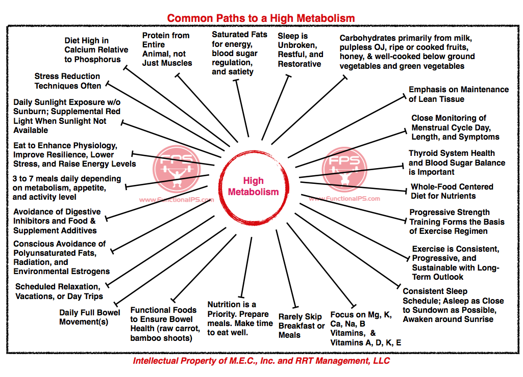
Dairy, Calcium, and Weight Management in Adults and Management
Belly Fat, Cortisol, and Stress
Stress and Aging: The Glucocorticoid Cascade Hypothesis

TNF-alpha and insulin resistance:

Trends Endocrinol Metab. 2000 Aug;11(6):212-7.
Potential role of TNF-alpha in the pathogenesis of insulin resistance and type 2 diabetes.d
Moller DE.
Tumor necrosis factor alpha (TNF-alpha) has well-described effects on lipid metabolism in the context of acute inflammation, as in sepsis. Recently, increased TNF-alpha production has been observed in adipose tissue derived from obese rodents or human subjects and TNF-alpha has been implicated as a causative factor in obesity-associated insulin resistance and the pathogenesis of type 2 diabetes. Thus, current evidence suggests that administration of exogenous TNF-alpha to animals can induce insulin resistance, whereas neutralization of TNF-alpha can improve insulin sensitivity. Importantly, results from knockout mice deficient in TNF-alpha or its receptors have suggested that TNF-alpha has a role in regulating in vivo insulin sensitivity. However, the absence of TNF-alpha action might only partially protect against obesity-induced insulin resistance in mice. Multiple mechanisms have been suggested to account for these metabolic effects of TNF-alpha. These include the downregulation of genes that are required for normal insulin action, direct effects on insulin signaling, induction of elevated free fatty acids via stimulation of lipolysis, and negative regulation of PPAR gamma, an important insulin-sensitizing nuclear receptor. Although current evidence suggests that neutralizing TNF-alpha in type 2 diabetic subjects is not sufficient to cause metabolic improvement, it is still probable that TNF-alpha is a contributing factor in common metabolic disturbances such as insulin resistance and dyslipidemia.

Nature. 1997 Oct 9;389(6651):610-4.
Protection from obesity-induced insulin resistance in mice lacking TNF-alpha function.
Uysal KT, Wiesbrock SM, Marino MW, Hotamisligil GS.
Obesity is highly associated with insulin resistance and is the biggest risk factor for non-insulin-dependent diabetes mellitus. The molecular basis of this common syndrome, however, is poorly understood. It has been suggested that tumour necrosis factor (TNF)-alpha is a candidate mediator of insulin resistance in obesity, as it is overexpressed in the adipose tissues of rodents and humans and it blocks the action of insulin in cultured cells and whole animals. To investigate the role of TNF-alpha in obesity and insulin resistance, we have generated obese mice with a targeted null mutation in the gene encoding TNF-alpha and those encoding the two receptors for TNF-alpha. The absence of TNF-alpha resulted in significantly improved insulin sensitivity in both diet-induced obesity and that resulting for the ob/ob model of obesity. The TNFalpha-deficient obese mice had lower levels of circulating free fatty acids, and were protected from the obesity-related reduction in the insulin receptor signalling in muscle and fat tissues. These results indicate that TNF-alpha is an important mediator of insulin resistance in obesity through its effects on several important sites of insulin action.

Nihon Rinsho. 1999 Mar;57(3):622-6.
[Syndrome X].
Kotake H, Oikawa S.
Insulin resistance is an early and major feature in the development of non-insulin-dependent diabetes mellitus (NIDDM). It is also associated with hyperlipidemia, hypertension, obesity and cardiovascular disease. It is the clustor of the risk factors for atherosclerosis and recognized as ‘insulin-resistance syndrome’ (Syndrome X). Central (abdominal) obesity is much more strongly associated with insulin resistance than overall obesity. The increase of both the influx of free fatty acid to liver and the production of TNF-alpha in adipose tissue may play an important role in mechanism of insulin resistance associated with central obesity. Calorie restriction and weight loss improve insulin sensitivity in overweight humans. Exercise training also improves insulin sensitivity via increased oxidative enzymes, glucose transporters (GLUT4) and capillarity in muscle as well as by reducing abdominal fat. The new ‘glitazones’ (thiazolidinediones) is used clinically to improve insulin sensitivity.

Physiol Res. 1998;47(4):215-25.
Thiazolidinediones–tools for the research of metabolic syndrome X.
Komers R, Vrána A.
“The resistance to insulin (insulin resistance, IR) is a common feature and a possible link between such frequent disorders as non-insulin dependent diabetes mellitus (NIDDM), hypertension and obesity…Furthermore, TDs inhibit the pathophysiological effects exerted by tumour-necrosis factor (TNF alpha), a cytokine involved in the pathogenesis of IR [insulin resistance].”

Exp Clin Endocrinol Diabetes. 1999;107(2):119-25.
Mechanisms of TNF-alpha-induced insulin resistance.
Hotamisligil GS.
There is now substantial evidence linking TNF-alpha to the presentation of insulin resistance in humans, animals and in vitro systems. We explored the relationship between TNF-alpha and insulin resistance using knockout mice deficient for either TNF-alpha or one or both of its receptors, p55 and p75. In studies of TNF-alpha-deficient knockout mice with diet-induced obesity, obese TNF-alpha knockouts responded to an exogenous dose of insulin or glucose much more efficiently than TNF-alpha wild-type animals. This finding suggests that deletion of TNF-alpha leads to increased insulin sensitivity, ie decreased insulin resistance. In studies using genetically obese ob/ob mice, TNF-alpha receptor wild-type and p75 receptor knockout animals developed a pronounced hyperinsulinemia and transient hyperglycaemia, whereas p55 receptor and double-knockout animals did not. Moreover, in glucose and insulin tolerance tests, we found that p75 knockout animals exhibited profiles identical to those of the wild-type animals, but that p55 knockout animals and double mutants showed a mild improvement in insulin sensitivity, relative to the wild type. Since the improvement in sensitivity was slightly greater with double mutants, p55 alone cannot be responsible for TNF-alpha’s promotion of insulin resistance in obese mice, despite the likelihood that it is more important than p75. How TNF-alpha-related insulin resistance is mediated is not fully clear, although phosphorylation of serine residues on IRS-1 has previously been shown to be important. When we monitored Glut 4 expression in obese TNF-alpha wild-type and knockout mice, we found no convincing evidence that TNF-alpha mediation of the down-regulation of Glut 4 mRNA expression is responsible for insulin resistance. However, we found an approximately 2-fold increase in insulin-stimulated tyrosine phosphorylation of the insulin receptor in the muscle and adipose tissue of TNF-alpha knockout mice, suggesting that insulin receptor signalling is an important target for TNF-alpha. Other possible mediators of TNF-alpha-induced insulin resistance include circulating free fatty acids (FFAs) and leptin.

TNF-alpha and obesity:

The Journal of Clinical Endocrinology & Metabolism August 1, 1998 vol. 83 no. 8 2907-2910
Tumor Necrosis Factor-α in Sera of Obese Patients: Fall with Weight Loss
Paresh Dandona, Ruth Weinstock, Kuldip Thusu, Ehad Abdel-Rahman, Ahmad Aljada, and Thomas Wadden
In view of the recent demonstration that obesity in animals and humans is associated with an increase in tumor necrosis factor-α (TNFα) expression, that this expression falls with weight loss, and that TNFα may specifically inhibit insulin action, the possibility that TNFα may be a mediator of insulin resistance has been raised. We have undertaken this study to investigate whether serum TNFα concentrations are elevated in obese subjects, whether they fall after weight loss, and whether this fall parallels the fall in insulin release after glucose challenge. Obese patients (age range: 25–54, weight mean ± sd: 96.4 ± 13.8 kg, body mass index: 35.7 ± 5.6 kg/m2) were started on a diet program. The mean weight fell to 84.5 ± 11.3 (P < 0.0001) and body mass index to 31.3 ± 4.9 (P < 0.0001). Plasma TNFα concentrations were markedly elevated in the obese (3.45 ± 0.16 pg/mL), when compared with controls (0.72 ± 0.28 pg/mL), and fell significantly (2.63 ± 1.40 pg/mL) after weight loss (P < 0.02). The magnitude of insulin release after glucose (75 g) challenge (area under the curve) also fell significantly (P < 0.01) after weight loss. The magnitude of weight loss and fall in TNFα were related to basal body weight (r = 0.57, P < 0.001) and basal TNFα (r = 0.55, P < 0.001) concentrations, respectively, but not to each other or to the glucose-induced insulin release (area under the curve). We conclude that obesity is associated with increased plasma TNFα concentrations, which fall with weight loss. Because circulating TNFα may mediate insulin resistance in the obese, a fall in TNFα concentrations may contribute to the restoration of insulin resistance after weight loss, Thus, TNFα may be an important circulating cytokine, which may provide a potentially reversible mechanism for mediating insulin resistance.

Endotoxin Increases TNF-alpha, Associated with Insulin Resistance and Diabetes:

Diabetes Care February 2011 vol. 34 no. 2 392-397
Endotoxemia Is Associated With an Increased Risk of Incident Diabetes
Pirkko J. Pussinen, PHD1, Aki S. Havulinna, MSC2, Markku Lehto, PHD3,4, Jouko Sundvall, MSC2 and Veikko Salomaa, MD2
OBJECTIVE Diabetes is accompanied with a chronic low-grade inflammation, which may in part be mediated by endotoxins derived from Gram-negative bacteria.
RESEARCH DESIGN AND METHODS We investigated in a population-based cohort whether endotoxemia is associated with clinically incident diabetes. The serum endotoxin activity was measured by limulus assay from the FINRISK97 cohort comprising 7,169 subjects aged 25–74 years and followed up for 10 years.
RESULTS Both the subjects with prevalent diabetes (n = 537) and those with incident diabetes (n = 462) had higher endotoxin activity than the nondiabetic individuals (P < 0.001). The endotoxin activity was significantly associated with increased risk for incident diabetes with a hazard ratio 1.004 (95% CI 1.001–1.007; P = 0.019) per unit increase resulting in a 52% increased risk (P = 0.013) in the highest quartile compared with the lowest one. The association was independent of diabetes risk factors: serum lipids, γ-glutamyl transferase, C-reactive protein, BMI, and blood glucose. Furthermore, the association of endotoxemia with an increased risk of incident diabetes was independent of the metabolic syndrome as defined either by the National Cholesterol Educational Program-Adult Treatment Panel III or the International Diabetes Federation. Endotoxin activity was linearly related (P < 0.001) to the number of components of the metabolic syndrome.
CONCLUSIONS Both prevalent and incident diabetes were associated with endotoxemia, which may link metabolic disorders to inflammation. The results suggest that microbes play a role in the pathogenesis of diabetes.

Diabetes Care. 2011 Feb;34(2):392-7.
Endotoxemia is associated with an increased risk of incident diabetes.
Pussinen PJ, Havulinna AS, Lehto M, Sundvall J, Salomaa V.
Both prevalent and incident diabetes were associated with endotoxemia, which may link metabolic disorders to inflammation. The results suggest that microbes play a role in the pathogenesis of diabetes.

Diabetes. 2007;56(7):1161-1772.
Metabolic Endotoxemia Initiates Obesity and Insulin Resistance
Patrice D. Cani; Jacques Amar; Miguel Angel Iglesias; Marjorie Poggi; Claude Knauf; Delphine Bastelica; Audrey M. Neyrinck; Francesca Fava; Kieran M. Tuohy; Chantal Chabo; Aurélie Waget; Evelyne Delmée; Béatrice Cousin; Thierry Sulpice; Bernard Chamontin; Jean Ferrières; Jean-François Tanti; Glenn R. Gibson; Louis Casteilla; Nathalie M. Delzenne; Marie Christine Alessi; Rémy Burcelin
Diabetes and obesity are two metabolic diseases characterized by insulin resistance and a low-grade inflammation. Seeking an inflammatory factor causative of the onset of insulin resistance, obesity, and diabetes, we have identified bacterial lipopolysaccharide (LPS) as a triggering factor. We found that normal endotoxemia increased or decreased during the fed or fasted state, respectively, on a nutritional basis and that a 4-week high-fat diet chronically increased plasma LPS concentration two to three times, a threshold that we have defined as metabolic endotoxemia. Importantly, a high-fat diet increased the proportion of an LPS-containing microbiota in the gut. When metabolic endotoxemia was induced for 4 weeks in mice through continuous subcutaneous infusion of LPS, fasted glycemia and insulinemia and whole-body, liver, and adipose tissue weight gain were increased to a similar extent as in high-fat-fed mice. In addition, adipose tissue F4/80-positive cells and markers of inflammation, and liver triglyceride content, were increased. Furthermore, liver, but not whole-body, insulin resistance was detected in LPS-infused mice. CD14 mutant mice resisted most of the LPS and high-fat diet-induced features of metabolic diseases. This new finding demonstrates that metabolic endotoxemia dysregulates the inflammatory tone and triggers body weight gain and diabetes. We conclude that the LPS/CD14 system sets the tone of insulin sensitivity and the onset of diabetes and obesity. Lowering plasma LPS concentration could be a potent strategy for the control of metabolic diseases.

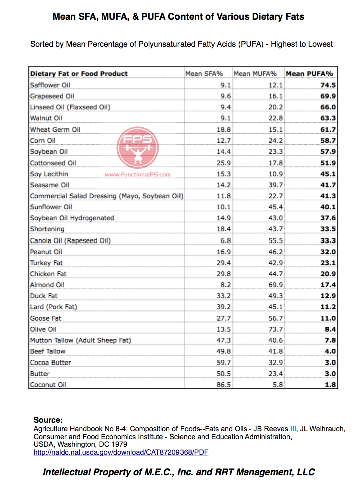
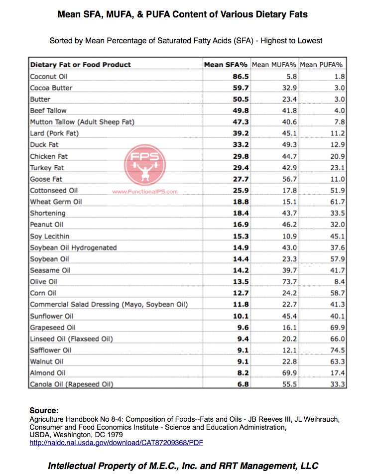
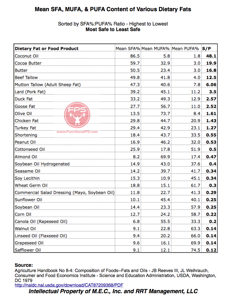
Saturated fats reduce endotoxemia, lipid peroxidation, and TNF-alpha:

Hepatology. 1997 Dec;26(6):1538-45.
Dietary saturated fatty acids down-regulate cyclooxygenase-2 and tumor necrosis factor alfa and reverse fibrosis in alcohol-induced liver disease in the rat.
Nanji AA, Zakim D, Rahemtulla A, Daly T, Miao L, Zhao S, Khwaja S, Tahan SR, Dannenberg AJ.
We investigated the potential of dietary saturated fatty acids to decrease endotoxemia and suppress expression of cyclooxygenase 2 (Cox-2) and tumor necrosis factor alpha (TNF-alpha) in established alcohol-induced liver injury. Six groups (five rats/group) of male Wistar rats were studied. Rats in group 1 were fed a fish oil-ethanol diet for 6 weeks. Rats in groups 2, 3, and 4 were fed fish oil and ethanol for 6 weeks. Ethanol administration was stopped at this time, and the rats were switched to isocaloric diets containing dextrose with fish oil (group 2), palm oil (group 3), or medium-chain triglycerides (group 4) as the source of fat for an additional 2 weeks. Rats in groups 5 and 6 were fed fish oil-ethanol and fish oil-dextrose, respectively, for 8 weeks. Liver samples were analyzed for histopathology, lipid peroxidation, and levels of messenger RNA (mRNA) for Cox-2 and TNF-alpha. Concentrations of endotoxin were determined in plasma. The most severe inflammation and fibrosis were detected in groups 1 and 5, as were the highest levels of endotoxin, lipid peroxidation, and mRNA for Cox-2 and TNF-alpha. After ethanol was discontinued, there was minimal histological improvement in group 2 but near normalization of the histology, including regression of fibrosis, in groups 3 and 4. Histological improvement was associated with decreased levels of endotoxin, lipid peroxidation, and reduced expression of Cox-2 and TNF-alpha. The data indicate that a diet enriched in saturated fatty acids (groups 3 and 4) effectively reverses alcohol-induced liver injury, including fibrosis. The therapeutic effects of saturated fatty acids may be explained, at least in part, by reduced endotoxemia and lipid peroxidation, which in turn result in decreased levels of TNF-alpha and Cox-2.

Lipoproteins protect against endotoxemia and TNF-alpha:

Eur Heart J. 1993 Dec;14 Suppl K:125-9.
The protective effect of serum lipoproteins against bacterial lipopolysaccharide.
Read TE, Harris HW, Grunfeld C, Feingold KR, Kane JP, Rapp JH.
Lipoproteins bind and inactivate bacterial endotoxin, both in vitro and in vivo. Both cholesterol ester-rich and TG-rich lipoproteins, and TG-rich lipid emulsions can prevent death in mice when pre-incubated with a lethal dose of endotoxin before intraperitoneal administration. Chylomicrons can also prevent death when given intravenously after endotoxin in rats. The metabolic fate of lipoprotein-bound endotoxin appears to be directed by the lipoprotein particle. When administered with chylomicrons, the plasma clearance and hepatic uptake of endotoxin are enhanced. Endotoxin is shunted preferentially to hepatocytes and away from hepatic macrophages, thereby increasing endotoxin excretion [corrected] in bile. The survival benefit and alterations in metabolism afforded by chylomicrons correlate with a reduction in peak serum levels of tumour necrosis factor (TNF), providing a possible mechanism by which lipoproteins protect against endotoxin-induced death. These findings suggest a possible role for lipoproteins or lipid emulsions in the body’s defence against endotoxaemia.

Dairy lowers TNF-alpha in the overweight:

Am J Clin Nutr January 2010 vol. 91 no. 1 16-22
Effects of dairy compared with soy on oxidative and inflammatory stress in overweight and obese subjects
Michael B Zemel, Xiaocun Sun, Teresa Sobhani, and Beth Wilson
Background: We recently showed that calcitriol increases oxidative and inflammatory stress; moreover, inhibition of calcitriol with high-calcium diets decreased both adipose tissue and systemic oxidative and inflammatory stress in obese mice, whereas dairy exerted a greater effect. However, these findings may be confounded by concomitant changes in adiposity.
Objective: The objective of this study was to evaluate the acute effects of a dairy-rich diet on oxidative and inflammatory stress in overweight and obese subjects in the absence of adiposity changes.
Design: Twenty subjects (10 obese, 10 overweight) participated in a blinded, randomized, crossover study of dairy- compared with soy-supplemented eucaloric diets. Two 28-d dietary periods were separated by a 28-d washout period. Inflammatory and oxidative stress biomarkers were measured on days 0, 7, and 28 of each dietary period.
Results: The dairy-supplemented diet resulted in significant suppression of oxidative stress (plasma malondialdehyde, 22%; 8-isoprostane-F2α, 12%; P < 0.0005) and lower inflammatory markers (tumor necrosis factor-α, 15%, P < 0.002; interleukin-6, 13%, P < 0.01; monocyte chemoattractant protein-1, 10%, P < 0.0006) and increased adiponectin (20%, P < 0.002), whereas the soy exerted no significant effect. These effects were evident by day 7 of treatment and increased in magnitude at the end of the 28-d treatment periods. There were no significant differences in response to treatment between overweight and obese subjects for any variable studied. Conclusion: An increase in dairy food intake produces significant and substantial suppression of the oxidative and inflammatory stress associated with overweight and obesity. This trial was registered at clinicaltrials.gov as NCT00686426.

Am J Clin Nutr August 2011 vol. 94 no. 2 422-430
Dairy attentuates oxidative and inflammatory stress in metabolic syndrome
Renée A Stancliffe, Teresa Thorpe, and Michael B Zemel
Background: Oxidative and inflammatory stress are elevated in obesity and are further augmented in metabolic syndrome. We showed previously that dairy components suppress the adipocyte- and macrophage-mediated generation of reactive oxygen species and inflammatory cytokines and systemic oxidative and inflammatory biomarkers in obesity.
Objective: The objective of this study was to determine the early (7 d) and sustained (4 and 12 wk) effects of adequate-dairy (AD) compared with low-dairy (LD) diets in subjects with metabolic syndrome.
Design: Forty overweight and obese adults with metabolic syndrome were randomly assigned to receive AD (3.5 daily servings) or LD (<0.5 daily servings) weight-maintenance diets for 12 wk. Oxidative and inflammatory biomarkers were assessed at 0, 1, 4, and 12 wk as primary outcomes; body weight and composition were measured at 0, 4, and 12 wk as secondary outcomes. Results: AD decreased malondialdehyde and oxidized LDL at 7 d (35% and 11%, respectively; P < 0.01), with further decreases by 12 wk. Inflammatory markers were suppressed with intake of AD, with decreases in tumor necrosis factor-α at 7 d and further reductions through 12 wk (35%; P < 0.05); decreases in interleukin-6 (21%; P < 0.02) and monocyte chemoattractant protein 1 (14% decrease at 4 wk, 24% decrease at 12 wk; P < 0.05); and a corresponding 55% increase in adiponectin at 12 wk (P < 0.01). LD exerted no effect on oxidative or inflammatory markers. Diet had no effect on body weight; however, AD significantly reduced waist circumference and trunk fat (P < 0.01 for both), and LD exerted no effect.
Conclusion: An increase in dairy intake attenuates oxidative and inflammatory stress in metabolic syndrome. This trial was registered at clinicaltrials.gov as NCT01266330.

Posted in General.

Tagged with , , , , , , , , , , , , , , , , , , , , , , , .


Leptin, Estrogen, Inflammation, Breast Cancer

Also see:
Sugar (Sucrose) Restrains the Stress Response
Endotoxin: Poisoning from the Inside Out
Ray Peat, PhD on Endotoxin
Hormonal profiles in women with breast cancer
Plasma Estrogen Does Not Reflect Tissue Concentration of Estrogen
Pre and Postmenopausal Women: Progesterone Decreases Aromatase Activity
Fat Tissue and Aging – Increased Estrogen
Estrogen Related to Loss of Fat Free Mass with Aging
How does estrogen enhance endotoxin toxicity? Let me count the ways.
Estrogen, Endotoxin, and Alcohol-Induced Liver Injury
Breast Cancer by Ray Peat
Preventing and treating cancer with progesterone by Ray Peat

Quotes by Ray Peat, PhD:
“I doubt that there is any biological significance in the idea of leptin resistance. Leptin promotes inflammation and cancer, so it might be good to be resistant to it, but I think the concept is mainly an outgrowth of the pharmaceutical industry’s promotion of leptin as a cure for obesity.”

“Leptin (which is promoted by estrogen) is a hormone produced by fat cells, and it, like estrogen, activates the POMC-related endorphin stress system. The endorphins activate histamine, another promoter of inflammation and cell division.

Progesterone opposes those various biochemical effects of estrogen in multiple ways, for example by inhibiting the ACTH stress response, by restraining cortisol’s harmful actions, and by inhibiting leptin.”

Cell Immunol. 2008 Mar-Apr;252(1-2):139-45. doi: 10.1016/j.cellimm.2007.09.004. Epub 2008 Mar 4.
Leptin beyond body weight regulation–current concepts concerning its role in immune function and inflammation.
Lago R, Gómez R, Lago F, Gómez-Reino J, Gualillo O.
Leptin, a 16 kDa non-glycosylated polypeptide produced primarily by adipocytes and released into the systemic circulation, exerts a multitude of regulatory functions including energy utilization and storage, regulation of various endocrine axes, bone metabolism, and thermoregulation. In addition to leptin’s best known role as regulator of energy homeostasis, several studies indicate that leptin plays a pivotal role in immune and inflammatory response. Because of its dual nature as a hormone and cytokine, leptin can be nowadays considered the link between neuroendocrine and immune system. The increase in leptin production that occurs during infections and inflammatory processes strongly suggests that this adipokine is a part of the cytokines network which governs inflammatory/immune response and host defence mechanisms. Indeed, leptin plays a relevant role in inflammatory processes involving either innate or adaptive immune responses. Several studies have implicated leptin in the pathogenesis of autoimmune inflammatory conditions such as encephalomyelitis, type I diabetes, bowel inflammation and also articular degenerative diseases such as rheumatoid arthritis and osteoarthritis. Although the mechanisms by which leptin exerts its action as modulator of inflammatory/immune response are likely to be more complex than predicted and far to be completely depicted, there is a general consensus about its pivotal role as pro-inflammatory and immune-modulating agent. Here, we review the most recent advances on leptin biology with a particular attention to its adipokine facet, even though its role as metabolic hormone will be also addressed.

Mol Cell Endocrinol. 2013 Apr 4. pii: S0303-7207(13)00121-4. doi: 10.1016/j.mce.2013.03.025. [Epub ahead of print]
Leptin-cytokine crosstalk in breast cancer.
Newman G, Gonzalez-Perez RR.
Despite accumulating evidence suggesting a positive correlation between leptin levels, obesity, post-menopause and breast cancer incidence, our current knowledge on the mechanisms involved in these relationships is still incomplete. Since the cloning of leptin in 1994 and its receptor (OB-R) 1 year later by Friedman’s laboratory (Zhang et al., 1994) and Tartaglia et al. (Tartaglia et al., 1995), respectively, more than 22,000 papers related to leptin functions in several biological systems have been published (Pubmed, 2012). The ob gene product, leptin, is an important circulating signal for the regulation of body weight. Additionally, leptin plays critical roles in the regulation of glucose homeostasis, reproduction, growth and the immune response. Supporting evidence for leptin roles in cancer has been shown in more than 1000 published papers, with almost 300 papers related to breast cancer (Pubmed, 2012). Specific leptin-induced signaling pathways are involved in the increased levels of inflammatory, mitogenic and pro-angiogenic factors in breast cancer. In obesity, a mild inflammatory condition, deregulated secretion of proinflammatory cytokines and adipokines such as IL-1, IL-6, TNF-α and leptin from adipose tissue, inflammatory and cancer cells could contribute to the onset and progression of cancer. We used an in silico software program, Pathway Studio 9, and found 4587 references citing these various interactions. Functional crosstalk between leptin, IL-1 and Notch signaling (NILCO) found in breast cancer cells could represent the integration of developmental, proinflammatory and pro-angiogenic signals critical for leptin-induced breast cancer cell proliferation/migration, tumor angiogenesis and breast cancer stem cells (BCSCs). Remarkably, the inhibition of leptin signaling via leptin peptide receptor antagonists (LPrAs) significantly reduced the establishment and growth of syngeneic, xenograft and carcinogen-induced breast cancer and, simultaneously decreased the levels of VEGF/VEGFR2, IL-1 and Notch. Inhibition of leptin-cytokine crosstalk might serve as a preventative or adjuvant measure to target breast cancer, particularly in obese women. This review is intended to present an update analysis of leptin actions in breast cancer, highlighting its crosstalk to inflammatory cytokines and growth factors essential for tumor development, angiogenesis and potential role in BCSC.

Pathol Oncol Res. 2006;12(2):69-72. Epub 2006 Jun 24.
Leptin–from regulation of fat metabolism to stimulation of breast cancer growth.
Sulkowska M, Golaszewska J, Wincewicz A, Koda M, Baltaziak M, Sulkowski S.
Leptin restricts intake of calories as a satiety hormone. It probably stimulates neoplastic proliferation in breast cancer, too. Growth of malignant cells could be regulated by various leptin-induced second messengers like STAT3 (signal transducers and activators of transcription 3), AP-1 (transcription activator protein 1), MAPK (mitogen-activated protein kinase) and ERKs (extracellular signal-regulated kinases). They seem to be involved in aromatase expression, generation of estrogens and activation of estrogen receptor alpha (ERalpha) in malignant breast epithelium. Leptin may maintain resistance to antiestrogen therapy. Namely, it increased activation of estrogen receptors, therefore, it was suspected to reduce or even overcome the inhibitory effect of tamoxifen on breast cell proliferation. Although several valuable reviews have been focused on the role of leptin in breast cancer, the status of knowledge in this field changes quickly and our insight should be continuously revised. In this summary, we provide refreshed interpretation of intensively reported scientific queries of the topic.

J Cell Biochem. 2008 Nov 1;105(4):956-64. doi: 10.1002/jcb.21911.
Leptin signaling in breast cancer: an overview.
Cirillo D, Rachiglio AM, la Montagna R, Giordano A, Normanno N.
The adipocyte-derived peptide leptin acts through binding to specific membrane receptors, of which six isoforms (obRa-f) have been identified up to now. Binding of leptin to its receptor induces activation of different signaling pathways, including the JAK/STAT, MAPK, IRS1, and SOCS3 signaling pathways. Since the circulating levels of leptin are elevated in obese individuals, and excess body weight has been shown to increase breast cancer risk in postmenopausal women, several studies addressed the role of leptin in breast cancer. Expression of leptin and its receptors has been demonstrated to occur in breast cancer cell lines and in human primary breast carcinoma. Leptin is able to induce the growth of breast cancer cells through activation of the Jak/STAT3, ERK1/2, and/or PI3K pathways, and can mediate angiogenesis by inducing the expression of vascular endothelial growth factor (VEGF). In addition, leptin induces transactivation of ErbB-2, and interacts in triple negative breast cancer cells with insulin like growth factor-1 (IGF-1) to transactivate the epidermal growth factor receptor (EGFR), thus promoting invasion and migration. Leptin can also affect the growth of estrogen receptor (ER)-positive breast cancer cells, by stimulating aromatase expression and thereby increasing estrogen levels through the aromatization of androgens, and by inducing MAPK-dependent activation of ER. Taken together, these findings suggest that the leptin system might play an important role in breast cancer pathogenesis and progression, and that it might represent a novel target for therapeutic intervention in breast cancer.

Mini Rev Med Chem. 2006 Aug;6(8):897-907.
Leptin, estrogens and cancer.
Maeso Fortuny MC, Brito Díaz B, Cabrera de León A.
Obesity is a state of leptin resistance in which the membrane leptin receptor and the JAK-STAT pathway are blocked. This leads to increased intracellular concentrations of lipid metabolites, increased non-oxidative metabolism by adipocytes, and stimulation of the cell estrogen cycle. These factors are potentially oncogenic via the shared mitogen-activated protein kinase (MAPK), mitogen/extracellular signal-regulated kinase (MEK) and extracellular signal-regulated kinase (ERK) cellular pathways.

Expert Opin Investig Drugs. 2005 Mar;14(3):251-64.
Parathyroid hormone and leptin–new peptides, expanding clinical prospects.
Whitfield JF.
Leptin, a member of the cytokine superfamily has a PTH-like osteogenic activity and may even partly mediate PTH action. But leptin has two drawbacks that cloud its therapeutic future. First, apart from directly stimulating osteoblastic cells, it targets cells in the hypothalamic ventromedial nuclei and through them it reduces oestrogenic activity by promoting osteoblast-suppressing adrenergic activity. Second, it stimulates vascular and heart valve ossification, which leads to such events as heart failure and diabetic limb amputations.

Endocrinology 2001 Jul;142(7):2796-804.
Sucrose ingestion normalizes central expression of corticotropin-releasing-factor messenger ribonucleic acid and energy balance in adrenalectomized rats: a glucocorticoid-metabolic-brain axis?
Laugero KD, Bell ME, Bhatnagar S, Soriano L, Dallman MF.
Both CRF and norepinephrine (NE) inhibit food intake and stimulate ACTH secretion and sympathetic outflow. CRF also increases anxiety; NE increases attention and cortical arousal. Adrenalectomy (ADX) changes CRF and NE activity in brain, increases ACTH secretion and sympathetic outflow and reduces food intake and weight gain; all of these effects are corrected by administration of adrenal steroids. Unexpectedly, we recently found that ADX rats drinking sucrose, but not saccharin, also have normal caloric intake, metabolism, and ACTH. Here, we show that ADX (but not sham-ADX) rats prefer to consume significantly more sucrose than saccharin. Voluntary ingestion of sucrose restores CRF and dopamine-beta-hydroxylase messenger RNA expression in brain, food intake, and caloric efficiency and fat deposition, circulating triglyceride, leptin, and insulin to normal. Our results suggest that the brains of ADX rats, cued by sucrose energy (but not by nonnutritive saccharin) maintain normal activity in systems that regulate neuroendocrine (hypothalamic-pituitary-adrenal), behavioral (feeding), and metabolic functions (fat deposition). We conclude that because sucrose ingestion, like glucocorticoid replacement, normalizes energetic and neuromodulatory effects of ADX, many of the actions of the steroids on the central nervous system under basal conditions may be indirect and mediated by signals that result from the metabolic effects of adrenal steroids.

Ginekol Pol. 1999 Jan;70(1):1-7.
Leptin regulation of aromatase activity in adipose stromal cells from regularly cycling women.
Magoffin DA, Weitsman SR, Aagarwal SK, Jakimiuk AJ.
OBJECTIVES AND DESIGN:
Leptin, a product of adipocytes, is a cytokine with multiple effects on the reproductive axis. Leptin causes the activation of STAT proteins within target cells. The aromatase gene promoter in adipose stromal cells contains a functional STAT binding region, leading to the hypothesis that leptin may regulate aromatase activity in fat tissue. To test this hypothesis, adipose stromal cells were isolated from subcutaneous abdominal fat or breast fat then placed into tissue culture.
MATERIALS AND METHODS:
The cells were treated for three days with increasing concentrations of recombinant human leptin. Aromatase activity in the stromal cells was measured by the release of 3H2O from radiolabeled androstenedione precursor.
RESULTS:
Basal aromatase activity varied markedly between, but there were no differences between abdominal fat and breast fat. Leptin concentrations in the physiological range of normal weight or thin women (10 ng/ml) had no effect on aromatase activity. In 2 of 8 abdominal fat cultures and 1 of 2 breast fat cultures, a high obese concentration of leptin (100 ng/ml) stimulated a significant increase in aromatase activity. In the remaining subjects there was no effect of leptin, even at high concentrations.
CONCLUSIONS:
These data demonstrate that in approximately 30 percent of our subject population leptin was able to stimulate aromatase activity in adipose stromal cells at high concentrations. The elevated levels of aromatase activity may contribute to increase circulating estrogen levels in certain obese women and suggest that elevated leptin concentrations in obese women may cause locally elevated estrogen concentrations in the breast and thereby promote tumor formation.

Contrib Nephrol. 2006;151:151-64.
Leptin as a proinflammatory cytokine.
Lord GM.
Leptin is a 16-kDa protein produced mainly by adipocytes. Animal models demonstrate that leptin is required for control of bodyweight and reproduction, since mice defective in leptin or the leptin receptor are obese, hyperphagic insulin resistant and infertile. Our initial series of observations lead us to propose that leptin also had significant effects on human type I proinflammatory immune responses. In support of this hypothesis, leptin deficient mice are resistant to a wide range of autoimmune diseases and display features of immune deficiency. Subsequent work has confirmed that leptin has a pleiotrophic role on the immune response and can rightly be considered, both structurally and functionally, as a proinflammatory cytokine.

Fertil Steril. 2010 Aug;94(3):1037-43.
Serum leptin levels, hormone levels, and hot flashes in midlife women.
Alexander C, Cochran CJ, Gallicchio L, Miller SR, Flaws JA, Zacur H.
To examine the associations between serum leptin levels, sex steroid hormone levels, and hot flashes in normal weight and obese midlife women.
DESIGN:
Cross-sectional study.
SETTING:
University clinic.
PATIENT(S):
201 Caucasian, nonsmoking women aged 45 to 54 years with a body mass index of <25 kg/m2 or >or=30 kg/m2.
INTERVENTION(S):
Questionnaire, fasting blood samples.
MAIN OUTCOME MEASURE(S):
Serum leptin and sex steroid hormone levels.
RESULT(S):
Correlation and regression models were performed to examine associations between leptin levels, hormone levels, and hot flashes. Leptin levels were associated with BMI, with “ever experiencing hot flashes” (questionnaire), with hot flashes within the last 30 days, and with duration of hot flashes (>1 year, P=.03). Leptin was positively correlated with testosterone, free testosterone index, and free estrogen index and inversely associated with levels of sex hormone-binding globulin. In women with a body mass index>or=30 kg/m2, leptin levels no longer correlated with testosterone levels.
CONCLUSION(S):
Serum leptin levels are associated with the occurrence and duration of hot flashes in midlife women; however, no correlation was found between leptin and serum estradiol.

Endocr Relat Cancer. 2010 Apr 21;17(2):373-82.
Cellular and molecular crosstalk between leptin receptor and estrogen receptor-{alpha} in breast cancer: molecular basis for a novel therapeutic setting.
Fusco R, Galgani M, Procaccini C, Franco R, Pirozzi G, Fucci L, Laccetti P,
Matarese G.
Obesity is associated with an increased risk of breast cancer. A number of adipocytokines are increased in obesity causing low-level chronic inflammation associated with an increased risk of tumors. The adipocytokine leptin shows profound anti-obesity and pro-inflammatory activities. We have hypothesized that in common obesity, high circulating leptin levels might contribute to an increased risk of breast cancer by affecting mammary cell proliferation and survival. Leptin exerts its activity not only through leptin receptor (LepR), but also through crosstalk with other signaling systems implicated in tumorigenesis. In this study, we focused our attention on the relationship between the leptin/LepR axis and the estrogen receptor-alpha (ERalpha). To this aim, we utilized two human breast cancer cell lines, one ERalpha-positive cell line (MCF 7) and the other ERalpha-negative cell line (MDA-MB 231). We observed that the two cell lines had a different sensitivity to recombinant leptin (rleptin): on MCF 7 cells, rleptin induced a strong phosphorylation of the signal transducer and activator of transcription (STAT) 3 and of the extracellular related kinase 1/2 pathways with an increased cell viability and proliferation associated with an increased expression of ERalpha receptor. This response was not present in the MDA-MB 231 cells. The effects induced by leptin were lost when LepR was neutralized using either a monoclonal inhibitory antibody to LepR or LepR gene-silencing siRNA. These data suggest that there is a bidirectional communication between LepR and ERalpha, and that neutralization and/or inactivation of LepR inhibits proliferation and viability of human breast cancer cell lines. This evidence was confirmed by ex vivo studies, in which we analyzed 33 patients with breast cancer at different stages of disease, and observed that there was a statistically significant correlation between the expression of LepR and ERalpha. In conclusion, this study suggests a crosstalk between LepR and ERalpha, and could envisage novel therapeutic settings aimed at targeting the LepR in breast cancers.

J Clin Endocrinol Metab. 2003 Mar;88(3):1285-91.
Endotoxin stimulates leptin in the human and nonhuman primate.
Landman RE, Puder JJ, Xiao E, Freda PU, Ferin M, Wardlaw SL.
Leptin, which plays a key role in regulating energy homeostasis, may also modulate the inflammatory response. An inflammatory challenge with endotoxin has been shown to stimulate leptin release in the rodent. This finding has not been reproduced in humans or in nonhuman primates, although leptin levels have been reported to increase in septic patients. We have therefore examined the effects of endotoxin injection on plasma leptin levels in nine ovariectomized monkeys and four postmenopausal women. In an initial study in five monkeys, mean leptin levels did not increase during the first 5 h after endotoxin treatment, but did increase significantly from 6.4 +/- 2.1 ng/ml at baseline to 12.3 +/- 4.4 ng/ml at 24 h (P = 0.043). In a second study, a significant increase in leptin over time was noted after endotoxin treatment (P < 0.001); leptin release during the 16- to 24-h period after endotoxin injection was 48% higher than during the control period (P = 0.043). A similar stimulatory effect of endotoxin on leptin was observed when monkeys received estradiol replacement. In a third study, repeated injections of endotoxin over a 3-d period stimulated IL-6, ACTH, cortisol, and leptin release (P < 0.001). Leptin increased during the first day of treatment in all animals, but only monkeys with baseline plasma leptin levels greater than 10 ng/ml exhibited a sustained increase in leptin throughout the 3-d period. There was a significant correlation (r = 0.81; P = 0.008) between the mean baseline leptin level and the percent increase in leptin over baseline on the last day of treatment. In the human subjects, plasma leptin concentrations did not change significantly during the 7-h period after endotoxin injection. However, leptin increased in all four women from a mean baseline of 8.34 +/- 3.1 to 13.1 +/- 4.3 ng/ml 24 h after endotoxin (P = 0.038). In summary, endotoxin stimulates the release of leptin into peripheral blood in the human and nonhuman primate, but the time course is different from that reported in the rodent. These results are consistent with previous reports of increased blood leptin levels in patients with sepsis. The significance of these findings and the potential role of leptin in modulating the response to inflammation in the human require further study.

J Leukoc Biol. 2000 Oct;68(4):437-46.
Leptin in the regulation of immunity, inflammation, and hematopoiesis.
Fantuzzi G, Faggioni R.
Leptin, the product of the ob gene, is a pleiotropic molecule that regulates food intake as well as metabolic and endocrine functions. Leptin also plays a regulatory role in immunity, inflammation, and hematopoiesis. Alterations in immune and inflammatory responses are present in leptin- or leptin-receptor-deficient animals, as well as during starvation and malnutrition, two conditions characterized by low levels of circulating leptin. Both leptin and its receptor share structural and functional similarities with the interleukin-6 family of cytokines. Leptin exerts proliferative and antiapoptotic activities in a variety of cell types, including T lymphocytes, leukemia cells, and hematopoietic progenitors. Leptin also affects cytokine production, the activation of monocytes/macrophages, wound healing, angiogenesis, and hematopoiesis. Moreover, leptin production is acutely increased during infection and inflammation. This review focuses on the role of leptin in the modulation of the innate immune response, inflammation, and hematopoiesis.

Exp Clin Endocrinol Diabetes. 1999;107(2):119-25.
Mechanisms of TNF-alpha-induced insulin resistance.
Hotamisligil GS.
There is now substantial evidence linking TNF-alpha to the presentation of insulin resistance in humans, animals and in vitro systems. We explored the relationship between TNF-alpha and insulin resistance using knockout mice deficient for either TNF-alpha or one or both of its receptors, p55 and p75. In studies of TNF-alpha-deficient knockout mice with diet-induced obesity, obese TNF-alpha knockouts responded to an exogenous dose of insulin or glucose much more efficiently than TNF-alpha wild-type animals. This finding suggests that deletion of TNF-alpha leads to increased insulin sensitivity, ie decreased insulin resistance. In studies using genetically obese ob/ob mice, TNF-alpha receptor wild-type and p75 receptor knockout animals developed a pronounced hyperinsulinemia and transient hyperglycaemia, whereas p55 receptor and double-knockout animals did not. Moreover, in glucose and insulin tolerance tests, we found that p75 knockout animals exhibited profiles identical to those of the wild-type animals, but that p55 knockout animals and double mutants showed a mild improvement in insulin sensitivity, relative to the wild type. Since the improvement in sensitivity was slightly greater with double mutants, p55 alone cannot be responsible for TNF-alpha’s promotion of insulin resistance in obese mice, despite the likelihood that it is more important than p75. How TNF-alpha-related insulin resistance is mediated is not fully clear, although phosphorylation of serine residues on IRS-1 has previously been shown to be important. When we monitored Glut 4 expression in obese TNF-alpha wild-type and knockout mice, we found no convincing evidence that TNF-alpha mediation of the down-regulation of Glut 4 mRNA expression is responsible for insulin resistance. However, we found an approximately 2-fold increase in insulin-stimulated tyrosine phosphorylation of the insulin receptor in the muscle and adipose tissue of TNF-alpha knockout mice, suggesting that insulin receptor signalling is an important target for TNF-alpha. Other possible mediators of TNF-alpha-induced insulin resistance include circulating free fatty acids (FFAs) and leptin.

Posted in General.

Tagged with , , , , , , , , , , , , , , , , , , , , , , , , , , , , , .


Common Paths to a High Metabolism

Also see:
Common Paths to a Low Metabolism
Promoters of Efficient v. Inefficient Metabolism
Collection of FPS Charts
Collection of Ray Peat Quote Blogs by FPS
Master List – Ray Peat, PhD Interviews
Components of Daily Energy Expenditure
Body Temperature, Metabolism, and Obesity

This chart provides several ways in which cell metabolism is optimized by nutrition and lifestyle factors. Multiple factors should be acting together. Juxtapose the chart in this blog with the Common Paths to a Low Metabolism chart.

Commonalities among the factors in the “Common Paths to a High Metabolism” chart are consistency in effort and preparation, lean tissue preservation or gain, focus on cellular needs, thyroid system support, blood sugar regulation, a diet rooted in historically relevant and digestible foodstuffs, emphasis on light exposure, recognition of the importance of regenerative sleep and a consistent sleep cycle, conscious avoidance of anti-metabolic factors, steroid hormone balance, and stress reduction.

FPS High Metabolism

Posted in General.

Tagged with , , , , , , .


Universal Principle of Cellular Energy

Also see:
Mitochondrial Medicine
Protect the Mitochondria
Collection of Ray Peat Quote Blogs by FPS
Carbon Dioxide as an Antioxidant
Carbon Dioxide Basics
Comparison: Carbon Dioxide v. Lactic Acid
Comparison: Oxidative Metabolism v. Glycolytic Metabolic
Promoters of Efficient v. Inefficient Metabolism
ATP Regulates Cell Water
Cardiolipin, Cytochrome Oxidase, Metabolism, & Aging
High Cholesterol and Metabolism
Low CO2 in Hypothyroidism
Protective Altitude
Lactate Paradox: High Altitude and Exercise
Protective Carbon Dioxide, Exercise, and Performance
Mitochondrial medicine
Low Carb Diet – Death to Metabolism
Low Blood Sugar Basics
The Cholesterol and Thyroid Connection
Thyroid Status and Oxidized LDL
Hypothyroidism and A Shift in Death Patterns
Light is Right
Using Sunlight to Sustain Life
PUFA Decrease Cellular Energy Production
PUFA Breakdown Products Depress Mitochondrial Respiration
“Curing” a High Metabolic Rate with Unsaturated Fats
Ray Peat, PhD on Carbon Dioxide, Longevity, and Regeneration
Altitude Improves T3 Levels
Mitochondria & Mortality
Altitude and Mortality
Lactate vs. CO2 in wounds, sickness, and aging; the other approach to cancer
Power Failure: Does mitochondrial dysfunction lie at the heart of common, complex diseases like cancer and autism?
Faulty Energy Production in Brain Cells Leads to Disorders Ranging from Parkinson’s to Intellectual Disability
Energy, structure, and carbon dioxide: A realistic view of the organism
Metabolic features of the cell danger response

When someone who has not been exposed to Ray Peat’s work, he/she can react with surprise when he/she hears that sugar, aspirin, milk/calcium, red light, salt, coffee, and saturated fats (to name a few) are recommended. This blog provides the foundational context required to begin to meaningfully interpret the 40+ years of writing from Dr. Peat.

The reason why the aforementioned therapies are important in a “Peatarian” lifestyle comes down to what I’m calling the Universal Principle of Cellular Energy but call the idea whatever you wish. The principle is universal in this way — it applies to all chronic health problems regardless of what medicine calls it. The thumbnail below attempts to provide the highlights of the concept.

UPCE fps

The substrate that cells need to make energy comes from the foods we eat; glucose oxidation produces the most ATP and carbon dioxide relative to other substrates and is synonymous with youthfulness and good health. This is why the dietary sugar component becomes important and carbohydrate avoidance is illogical.

Cellular energy deficiency leads to decreased renewal and the rate of aging speeds up progressively as adaptation becomes increasingly imperfect as cells become exhausted. If the energy crisis is severe, it can lead to cell death. Aging and disease(s) are representations of the body’s unique series of adaptations to an energy problem. The needs of your cells guide what actions your physiology takes to survive. Your cells can only do what the environment allows so it’s up to us to provide the optimal environment for energy success.

The principle can serve as the base by which nutritional decisions or therapies can be implemented. It’s also helpful in recognizing where someone may have done harm in his/her past. If you’re in a health rut, start by recognizing factors that slow down energy production and learn how to oppose these factors with pro-energy foods, lifestyle change, and supplements. Chief among the anti-energy factors are estrogen, polyunsaturated fats, endotoxin, serotonin, nitric oxide, darkness, and radiation. Thanks to inspiration by Dr. Peat, the FPS blog provides information on all of these factors.

Supportive Quotes by Ray Peat, PhD:
“If we learn to see problems in terms of a general disorder of energy metabolism, we can begin to solve them.”

“A given structure makes possible a certain level of useful energy, and adequate energy makes possible the maintenance of structure, and the advance to a higher and more efficient structural level.”

“I started my work with progesterone and related hormones in 1968. In papers in Physiological Chemistry and Physics (1971 and 1972) and in my dissertation (University of Oregon, 1972), I outlined my ideas regarding progesterone, and the hormones closely related to it, as protectors of the body’s structure and energy against the harmful effects of estrogen, radiation, stress, and lack of oxygen.

The key idea was that energy and structure are interdependent, at every level.

Since then, I have been working on both practical and theoretical aspects of this view. I think only a new perspective on the nature of living matter will make it possible to properly take advantage of the multitude of practical and therapeutic effects of the various life-supporting substances–pregnenolone, progesterone, thyroid hormone, and coconut oil in particular.”

“Life interposes itself between the “poles” of energy flow, and the flowing energy creates organization and structure, as it is dissipated into heat. Structures store some of the energy, and tend to increase in complexity, taking advantage of the flow of energy to create phase differences with expanded internal surfaces, like a finely mixed emulsion. Like a finely divided emulsion, the more highly energized the organism is, the stabler it is.”

“It seems that all of the problems of development and degeneration can be alleviated by the appropriate use of the energy-protective materials. When we realize that our human nature is problematic, we can begin to explore our best potentials.”

“A high level of respiratory energy production that characterizes young life is needed for tissue renewal. The accumulation of factors that impair mitochondrial respiration leads to increasing production of stress factors, that are needed for survival when the organism isn’t able to simply produce energetic new tissue as needed. Continually resorting to these substances progressively reshapes the organism, but the investment in short-term survival, without eliminating the problematic factors, tends to exacerbate the basic energy problem.”

“The biological idea of stress refers to the difficulty of adapting, and this involves energy, structure, and insight/orientation. Given enough energy, we can often adjust our structure to achieve full adaptation, and with insight, we can minimize the amount of energy and structural change needed, for example just by a change of pace or rhythm.”

“The metabolic interpretation of disease that had been making progress for several decades was suddenly submerged when government research financing began concentrating on genetic and viral interpretations of disease.”

“The availability of energy is central to our stable functioning, and the need for energy powerfully modifies our functioning.”

“Stress is an energy problem, that leads to the series of hormonal and metabolic reactions –lipolysis, glycolysis, increased serotonin, cortisol, estrogen, prolactin, leaky capillaries, protein catabolism, etc.”

“There is a repolarization of the cell after the production of ATP and CO2; the cell then gets ready to make more energy. Essentially the cell should be in a relaxed state as it gathers all of its forces to make energy. It then makes energy and goes back into its readiness state.”

“The intensity of oxidative metabolism is the basic factor that permits continuing coordination of activity, and harmonious renewal of all the components of the organism.”

“If we optimize the known factors which improve energy production (red light, short-chain and medium-chain saturated fats, and pregnenolone, for example), to the extent that our metabolism resembles that of a ten year old child, I don’t think there is any reason to suppose that we wouldn’t have regenerative, healing abilities which are common at that age.”

“In every type of tissue, it is the failure to oxidize glucose that produces oxidative stress and cellular damage.”

“Energy metabolism is the central biochemical issue.”

“Energy generates order and maintains it. Destruction of order degrades the ability of cells to produce energy.”

“Metabolic energy is fundamental to the development and maintenance of the body, and to the “ways in which living beings react to changed circumstances.” It’s an obvious first thing to consider when thinking about any “disease,” whether it’s cancer, radiation, sickness, dementia, depression, or traumatic injury.”

“The regulation of cell renewal probably involves all of the processes of life, but there are a few simple, interacting factors that suppress renewal. The accumulation of polyunsaturated fats, interacting with a high concentration of oxygen, damages mitochondria, and causes a chronic excessive exposure to cortisol. With mitochondrial damage, cells are unable to produce the progesterone needed to oppose cortisol and to protect cells.

Choosing the right foods, the right atmosphere, the right mental and physical activities, and finding the optimal rhythms of light, darkness, and activity, can begin to alter the streaming renewal of cells in all the organs. Designing a more perfect environment is going to be much simpler than the schemes of the genetic engineers. “

“Aging is an energy problem, and in the brain, which has extremely high energy requirements, interference with the energy supply quickly causes cells to die.”

“The balance between what a tissue needs and what it gets will govern the way that tissue functions, in both the short term and the long term. When a cell emits lactic acid and free radicals and the products of lipid peroxidation, it’s reasonable to assume that it isn’t getting everything that it needs, such as oxygen and glucose. With time, the cell will either die or adapt in some way to its deprived conditions.”

“The needs on the cellular level guide the organism’s adaptation.”

“Many of the anti-adaptive features of old blood can be reduced. Long daylight hours and high altitude (the altitude “lactate paradox” is an example of a cellular oxidative increase caused by lower oxygen pressure) shift the balance of some of the factors, and others can be improved by modifying the diet, and supplementing with things such as the protective steroids, thyroid hormone, aspirin, niacinamide (which can increase the oxidized state of NADH/NAD+), and caffeine. Providing the things needed for cellular energy while blocking some of the maladaptive factors approaches the problem of aging in a fundamental and holistic way.”

“In the excessively sensitive condition produced by hypoglycemia, several things happen that contribute to the maladaptive exaggerated inflammatory response. Adrenaline increases in hypoglycemia, and, if the adrenaline fails to convert glycogen into glucose, it will provide an alternative fuel by liberating free fatty acids from fat cells. If the liberated fatty acids are unsaturated, they will cause serotonin to be secreted, and both serotonin and the unsaturated fatty acids will suppress mitochondrial respiration, exacerbating the hypoglycemia. They will stimulate the release of cytokines, activating a variety of immunological and inflammatory processes, and they will cause blood vessels to become leaky, creating edema and starting the first stages of fibrosis. Both adrenaline and serotonin will stimulate the release of cortisol, which mobilizes amino acids from tissues such as the large skeletal muscles. Those muscles contain a large amount of cysteine and tryptophan, which, among other effects, suppress the thyroid. The increased tryptophan, especially in the presence of free fatty acids, is likely to be converted into additional serotonin, since fatty acids release tryptophan from albumin, increasing its entry into the brain. Free fatty acids and increased serotonin reduce metabolic efficiency (leading to insulin resistance, for example) and promote an inflammatory state.”

“When the available energy doesn’t meet the cell’s energy requirements, if the cell isn’t quickly killed by stress, it will use some adaptive mechanisms, stopping some repair processes to reduce energy expenditure, possibly stopping specialized functions to reduce energy needs. Fibrotic changes occur as a result of defensive reactions in stressed cells, usually following long periods of fatigue and inflammation.”

“When cells are in an energy deficient state, as in hypothyroidism, they are in the leaky edematous state.”

“Szent-Gyorgyi observed that, although ATP was involved in the contractions of muscles, its post-mortem disappearance caused the contraction and hardening of muscle known as rigor mortis. When he put hardened dead muscles into a solution of ATP, they relaxed and softened. The relaxed state is a state with adequate energy reserves.”

“Cells in their excited and exhausted state are increasingly open to penetration of toxins because of their own increased permeability and because of the increased leakiness of the blood vessels. Certain environmental toxins accumulate more rapidly as the cells lose their ability to destroy them. Several kinds of toxins, including unsaturated fats, inhibit the proteolytic enzymes that remodel tissue, and reduce the ability to dismantle and rebuild the cellular matrix.”

“Sugar can be used to produce energy with or without oxygen, but oxidative metabolism is about 15 times more efficient than the non-oxidative “glycolytic” or fermentive metabolism; higher organisms depend on this high efficiency oxidation for maintaining integration and normal functioning: If there is a small interference with respiration, the organism can adapt by increasing the rate of glycolysis, but there must be enough sugar to meet the demand. A response to stimulation is the production of more energy, with a proportional increase of oxygen and sugar consumption by the stimulated tissue; this produces more carbon dioxide. which enlarges the blood vessels in the area, providing more sugar and oxygen. If the irritation becomes destructive, efficiency is lost: oxygen is either consumed wastefully, causing blueness of the tissue (assuming circulation continues: blueness can also indicate bad circulation), or is not consumed. causing redness of the tissue. As more sugar is consumed in compensation, lactic acid also enlarges the blood vessels.

If the inflamed or exhausted tissue is small, the lactic acid can be consumed by other oxidizing tissues, sufficient sugar usually can be supplied, and repair occurs. But a large inflammation. or profound exhaustion, will lower the blood sugar systemically, and will deliver large amounts of lactic acid to the liver. The liver synthesizes glucose from the lactic acid, but at the expense of about 6 times more energy than is obtained from the inefficient metabolism – so that organismically, that tissue becomes 90 times less efficient than its original state. Besides this, an idle destruction of energy molecules (ATP or creatine phosphate) will increase the wastefulness even more.”

“The loss of control over water in the body is a result of energy failure…”

“As in other cells, ATP maintains the proper water content of cells.”

“…the essential element of stress is the inadequacy of energy to meet a challenge, and when energy is inefficient water is taken up.”

“When respiration is blocked tissues take up water.”

“Stress increases metabolic rate in a destructive, age accelerating way, with increased inflammation, and decreased resting oxidative metabolic rate. It’s the basic metabolic rate, with fast nerve conduction, quick cellular adaptation, etc., that’s biologically valuable.”

“The result of these passive and active processes is that each kind of ion has a characteristic concentration in each compartment, according to the metabolic energy state of the organism. 

Magnesium and potassium are mainly intracellular ions, sodium and calcium are mainly extracellular ions. When cells are excited, stressed, or de-energized, they lose magnesium and potassium, and take up sodium and calcium. The mitochondria can bind a certain amount of calcium during stress, but accumulating calcium can reach a point at which it inactivates the mitochondria, forcing cells to increase their inefficient glycolytic energy production, producing an excess of lactic acid. Abnormal calcification begins in the mitochondria. 

When cells are stressed or dying, they take up calcium, which tends to excite the cells at the same time that it inhibits their energy production, intensifying their stress. A cramp or a seizure is an example of uncontrolled cellular excitation. Prolonged excitation and stress contribute to tissue inflammation and fibrosis.

Gross calcification generally follows the fibrosis that is produced by inflammation.

Arteries, kidneys, and other organs calcify during aging. At the age of 90, the amount of calcium in the elastic layer of an artery is about 35 times greater than at the age of 20. Nearly every type of tissue, including the brain, is susceptible to the inflammatory process that leads through fibrosis to calcification. The exception is the skeleton, which loses its calcium as the soft tissues absorb calcium.

These observations lead to some simplifying ideas about the nature of aging and disease.

Some people who know about the involvement of calcium in aging, stress, and degeneration suggest eating a low calcium diet, but since we all have skeletons, dietary calcium restriction cant protect our cells, and in fact, it usually intensifies the process of calcification of the soft tissues. Statistics from several countries have clearly shown that the mortality rate (especially from arteriosclerotic heart disease, but also from some other diseases, including cancer) is lower than average in regions that have hard water, which often contains a very large amount of either calcium or magnesium.”

“Much of the intracellular magnesium is complexed with ATP, and helps to stabilize that molecule. If cellular energy production is low, as in hypothyroidism, cells tend to lose their magnesium very easily, shifting the balance toward the lower energy molecule, ADP, with the release of phosphate. ADP complexes with calcium, rather than magnesium, increasing the cells calcium content.”

“Degenerative diseases, especially cancer, heart disease, and brain diseases, are less prevalent in populations that live at a high altitude. When oxygen pressure is low, the lungs lose carbon dioxide more slowly, and so the amount of carbon dioxide retained in the body is greater. If the basic problem in hypothyroidism is the deficient production of carbon dioxide causing excessive loss of salt and retention of water, resulting in hypo-osmotic body fluids, then we would expect people at high altitude to have better retention of salt, more loss of water, and more hypertonic body fluids.”

“Since respiratory metabolism, governed by the thyroid hormone, is our main source of carbon dioxide, it’s obvious that thyroid deficiency should impair our ability to regulate water and solutes, such as salt.”

“The degenerative diseases can be seen as the cumulative result of stress, in which tissue damage results from the diabetes-like impairment of energy production.”

“Any stress or energy deficit that disturbs cellular structure or function disturbs the interactions among water, proteins, and other components of the cell. Excitation causes a cell to take up extra water, not by osmosis resulting from an increase in the concentration of solutes in the cell, or because the membrane has become porous, but because the structural proteins of the cell have momentarily increased their affinity for water.

This increased affinity is similar to the process that causes a gel to swell in the presence of alkalinity, and it is related to the process called electroosmosis, in which water moves toward a higher negative charge. Intense excitation or stress increases the cell’s electrically negative charges, and causes it to become more alkaline and to swell. Swelling and alkalinity cause the cell to begin the synthesis of DNA, in preparation for cell division.”

“The higher rate of metabolism produced by adequate thyroid function maintains a high rate of renewal of the cell’s systems, keeping the cell constantly adjusted to slight changes in the organism’s needs.”

“In hypothyroidism and diabetes, respiration is impaired, and lactic acid is formed even at rest, and relatively little carbon dioxide is produced. To compensate for the metabolic inefficiency of hypothyroidism, adrenalin and noradrenalin are secreted in very large amounts. Adrenalin causes free fatty acids to circulate at much higher levels, and the lactic acid, adrenalin, and free fatty acids all stimulate hyperventilation. The already deficient carbon dioxide is reduced even more, producing respiratory alkalosis. Free fatty acids, especially unsaturated fats, increase permeability of blood vessels, allowing proteins and fats to enter the endothelium and smooth muscle cells of the blood vessels. Lactic acid itself promotes an inflammatory state, and in combination with reduced CO2 and respiratory alkalosis, contributes to the hyponatremia (sodium deficiency) that is characteristic of hypothyroidism. This sodium deficiency and osmotic dilution causes cells to take up water, increasing their volume.”

“Thyroid, which opposes estrogen’s effects on cell energy, stimulates oxidative metabolism with the production of carbon dioxide, and reduces the water content of tissues.”

“Estrogen seems to work by blocking oxidative metabolism, and its first visible effect is to cause the stimulated tissue to take up water. Anything that causes cells to take up water seems to stimulate cell division.”

“This is where the issue of cell water comes in. Carbon dioxide, produced by oxidative cell metabolism, is associated with the high energy state of the cell. When something interferes with oxidative metabolism, lactic acid is produced instead of carbon dioxide. If the cell stays very long in this low oxygen state, it swells, taking up water. (The fatigued muscle, for example, can take up so much water in a short time that it weighs 20% more than before it began working so intensely that its energy needs far exceeded the availability of oxygen. This swelling is what causes the soreness and tightness of intense exercise. The swelling persists long after the liver has cleared the lactic acid from the blood.) This swelling from taking up water is involved in one type of “edema,” and in inflammation, or activation of the cells by hormones, as well as by simple oxygen deprivation.”

“Lactate formation from glucose is increased when anything interferes with respiratory energy production, but lactate, through a variety of mechanisms, can itself suppress cellular respiration. (This has been called the Crabtree effect.) Lactate can also inhibit its own formation, slowing glycolysis. In the healthy cell, the mitochondrion keeps glycolysis working by consuming pyruvate and electrons (or “hydrogens”) from NADH, keeping the cell highly oxidized, with a ratio of NAD+/NADH of about 200. When the mitochondrion’s ability to consume pyruvate and NADH is limited, the pyruvate itself accepts the hydrogen from NADH, forming lactic acid and NAD+ in the process. As long as lactate leaves the cell as fast as it forms, glycolysis will provide ATP to allow the cell to survive. Oxygen and pyruvate are normally “electron sinks,” regenerating the NAD+ needed to produce energy from glucose.

But if too much lactate is present, slowing glycolytic production of ATP, the cell with defective respiration will die unless an alternative electron sink is available. The synthesis of fatty acids is such a sink, if electrons (hydrogens) can be transferred from NADH to NADP+, forming NADPH, which is the reducing substance required for turning carbohydrates and pyruvate and amino acids into fats.”

“While Warburg was investigating the roles of glycolysis and respiration in cancer, a physician with a background in chemistry, W.F. Koch, in Detroit, was showing that the ability to use oxygen made the difference between health and sickness, and that the cancer metabolism could be corrected by restoring the efficient use of oxygen. He argued that a respiratory defect was responsible for immunodeficiency, allergy, and defective function of muscles, nerves, and secretory cells, as well as cancer. Koch’s idea of cancer’s metabolic cause and its curability directly challenged the doctrine of the genetic irreversibility of cancer that was central to governmental and commercial medical commitments.”

“A focus on correcting the respiratory defect would be relevant for all of the diseases and conditions (including heart disease, diabetes, dementia) involving inflammation and inappropriate excitation, not just for cancer.”

“Aging is characterized by loss of lean body mass, immunodeficiency, and a variety of autoimmune reactions. My perennial argument has been that decreased thyroid and progesterone, associated with increased estrogen and stress hormones, are largely responsible for those changes.”

“When respiratory energy production is blocked in stimulated cells, the cells are likely to die. (Cortisol, estrogen, polyunsaturated oils have this effect, especially on thymus cells.)”

“Thyroid is needed to keep the cell in an oxidative, rather than reductive state, and progesterone (which is produced elsewhere only when cells are in a rapidly oxidizing state) activates the processes that remove estrogen from the cell, and inactivates the processes that would form new estrogen in the cell.

Thyroid, and the carbon dioxide it produces, prevent the formation of the toxic lactic acid. When there is enough carbon dioxide in the tissues, the cell is kept in an oxidative state, and the formation of toxic free radicals is suppressed. Carbon dioxide therapy is extremely safe.”

“The organism can only be understood in its environments, and a cell can’t be understood without reference to the tissue and organism in which it lives. Although the geneticists were at first hostile to the idea that nutrition and geography could have anything to do with cancer, they soon tried to dominate those fields, insisting that mutagens and ethnicity would explain everything. But the evidence now makes it very clear that environment and nutrition affect the risk of cancer in ways that are not primarily genetic.”

“Substances such as PTH, nitric oxide, serotonin, cortisol, aldosterone, estrogen, thyroid stimulating hormone, and prolactin have regulatory and adaptive functions that are essential, but that ideally should act only intermittently, producing changes that are needed momentarily. When the environment is too stressful, or when nutrition isn’t adequate, the organism may be unable to mobilize the opposing and complementary substances to stop their actions. In those situations, it can be therapeutic to use some of the nutrients as supplements.”

“The movement of substances from blood to cell, and from cell to cell, is normally very tightly controlled, and when the systems that control those movements of water and its solutes are damaged, the tissues’ structures and functions are altered. The prevention of inappropriate leakiness can protect against the degenerative processes, and against aging itself, which is, among other things, a state of generalized leakiness.

When cells’ energy is depleted, water and various dissolved molecules are allowed to move into the cells, out of the cells, and through or around cells inappropriately. The weakened cells can even permit whole bacteria and similar particles to pass into and out of the blood stream more easily.

One of the earliest investigators of the effects of stress and fatigue on nerves and other cells was A.P. Nasonov, in the first half of the 20th century. A.S. Troshin (1956) has reviewed his work in detail. He showed that in cells as different as algae and nerve cells, fatigue caused them to take up dyes, and that the dyes were extruded, if the cells were able to recover their energy. When nerve cells are excited for a fraction of a second, they take up sodium and calcium, but quickly eliminate them. Prolonged excitation, leading to fatigue, can gradually shift the balance, allowing more substances to enter, and to stay longer.”

“If the cancer-productive field is taken into account, all of the factors that promote and sustain that field should be considered during therapy.

Two ubiquitous carcinogenic factors that can be manipulated without toxins are the polyunsaturated fatty acids (PUFA) and estrogen. These closely interact with each other, and there are many ways in which they can be modulated.

For example, keeping cells in a well oxygenated state with thyroid hormone and carbon dioxide will shift the balance from estradiol toward the weaker estrone. The thyroid stimulation will cause the liver to excrete estrogen more quickly, and will help to prevent the formation of aromatase in the tissues. Low temperature is one of the factors that increases the formation of estrogen. Lactic acid, serotonin, nitric oxide, prostaglandins, and the endorphins will be decreased by the shift toward efficient oxidative metabolism.

Progesterone synthesis will be increased by the higher metabolic rate, and will tend to keep the temperature higher.

Thyroid hormone, by causing a shift away from estrogen and serotonin, lowers prolactin, which is involved in the promotion of several kinds of cancer.

Vitamin D and vitamin K have some antiestrogenic effects. Vitamin D and calcium lower the inflammation-promoting parathyroid hormone (PTH).

Eliminating polyunsaturated fats from the diet is essential if the bystander effect is eventually to be restrained. Aspirin and salicylic acid can block many of the carcinogenic effects of the PUFA. Saturated fats have a variety of antiinflammatory and anticancer actions. Some of those effects are direct, others are the result of blocking the toxic effects of the PUFA. Keeping the stored unsaturated fats from circulating in the blood is helpful, since it takes years to eliminate them from the tissues after the diet has changed. Niacinamide inhibits lipolysis. Avoiding over-production of lipolytic adrenaline requires adequate thyroid hormone, and the adjustment of the diet to minimize fluctuations of blood sugar.”

“Failure to renew cells and tissues leads to loss of function and substance. Bones and muscles get weaker and smaller with aging. Diminished bone substance, osteopenia, is paralleled, at roughly the same rate, by the progressive loss of muscle mass, sarcopenia (or myopenia). The structure of aging tissue changes, with collagen tending to fill the spaces left by the disappearing cells. It’s also common for fat cells to increase, as muscle cells disappear.”

“Excitotoxicity, in its simplest sense, is the harmful cellular effect (death or injury) caused by an excitatory transmitter such as glutamate or aspartate acting on a cell whose energetic reserves aren’t adequate to sustain the level of activity provoked by the transmitter. Once an excitotoxic state exists, the consequences of cell exhaustion can increase the likelihood that the condition will spread to other cells, since any excitation can trigger a complex of other excitatory processes. As calcium enters cells, potassium leaves, and enzymes are activated, producing free fatty acids (linoleic and arachidonic, for example) and prostaglandins,”

“Simply getting outside the world of compartmentalized diseases, there is an abundance of evidence showing the variety of ways in which cells can fail. Energy is needed for cell maintenance and adaptation, and the type of fuel used to provide the energy is crucial. Fatty acids interfere with the oxidation of glucose, and this effect can be seen in heart failure, immunodeficiency, dementia, as well as in simple stress, diabetes, and many other simple situations (dementia: Montine and Morrow, 2005; Yaqoob, et al., 1994).”

“When a muscle or nerve is fatigued, it swells, retaining water. When the swelling is extreme, its ability to contract is limited. Excess water content resembles a partly excited state, in which increase amounts of sodium and calcium are free in the cytoplasm. Energy is needed to eliminate the sodium and calcium, or to bind calcium allowing the cell to extrude excess water and return to the resting state. Thyroid hormone allows cells’ mitochondria to efficiently produce energy, and it also regulates the synthesis of proteins (phospholamban and calcisequestrin) that control the binding of calcium. When the cell is energized, by the mitochondria working with thyroid, oxygen, and sugar, these proteins change their form, binding calcium and removing it from the contractile system, allowing the cell to relax, to be fully prepared for the next contraction. If the calcium isn’t fully and quickly bound, the cell retains extra water and sodium, and isn’t able to fully relax.”

“Older ways of understanding aging and degenerative disease are now returning to the foreground. The developmental interactions of the organism with its environment, and the interactions of its cells, tissues, and organs with each other, have again become the focus of biological aging research. In place of the old belief that “we are defined and limited by our genes,” the new perspective is showing us that we are limited by our environment, and that our environment can be modified. As we react to unsuitable environments, our internal environments become limiting for our cells, and instead of renewing themselves, repairing damage, and preparing for new challenges, our cells find themselves in blind alleys. Looking at aging in this way suggests that putting ourselves into the right environments could prevent aging.”

The end product of respiration is carbon dioxide, and it is an essential component of the life process. The ability to produce and retain enough carbon dioxide is as important for longevity as the ability to conserve enough heat to allow chemical reactions to occur as needed.

Carbon dioxide protects cells in many ways. By bonding to amino groups, it can inhibit the glycation of proteins during oxidative stress, and it can limit the formation of free radicals in the blood; inhibition of xanthine oxidase is one mechanism (Shibata, et al., 1998). It can reduce inflammation caused by endotoxin/LPS, by lowering the formation of tumor necrosis factor, IL-8 and other promoters of inflammation (Shimotakahara, et al., 2008). It protects mitochondria (Lavani, et al., 2007), maintaining (or even increasing) their ability to respire during stress.

The “replicative lifespan” of a cell can be shortened by factors like resveratrol or estrogen that interfere with mitochondrial production of carbon dioxide. Both of those chemicals cause skin cells, keratinocytes, to stop dividing, to take up calcium, and to begin producing the horny material keratin, that allows superficial skin cells to form an effective barrier. This process normally occurs as these cells differentiate from the basal (stem) cells and, by multiplying, move farther outward away from the underlying blood vessels that provide the nutrients that are oxidized to form carbon dioxide, and as they get farther from the blood supply, they get closer to the external air, which contains less than 1% as much CO2 as the blood. This normally causes their eventual hardening into the keratin cells, but when conditions are optimal, numerous layers of moist, translucent cells that give the skin the characteristic appearance of youth, will be retained between the basal cells and the condensed surface layers. (Wilke, et al., 1988)

In other types of tissue, a high level of carbon dioxide has a similar stabilizing effect on cells, preserving stem cells, limiting stress and preventing loss of function. In the lining of the mouth, where the oxygen tension is lower, and carbon dioxide higher, the cells don’t form as much keratin as the skin cells do. In the uterus, the lining cells would behave similarly, except that estrogen stimulates keratinization. A vitamin A deficiency mimics an estrogen excess, and can cause excessive keratinization of membrane cells.”

“Apparently, anything that depletes the cell’s energy, lowering ATP, allows an excess of calcium to enter cells, contributing to their death (Ray, et al., 1994). Increasing intracellular calcium activates phospholipases, releasing more polyunsaturated fats (Sweetman, et al., 1995) The acrolein which is released during lipid peroxidation inhibits mitochondrial function by poisoning the crucial respiratory enzyme, cytochrome oxidase, resulting in a decreased ability to produce energy (Picklo and Montine, 2001). (In the retina, the PUFA contribute to light-induced damage of the energy producing ability of the cells [King, 2004], by damaging the same crucial enzyme.)”

Posted in General.

Tagged with , , , , , , , , , , , , , , , , , , , , , , , , , , , , , , , , , , , , .


Inflammatory C-Reactive Protein (CRP)

Also see:
Endotoxin: Poisoning from the Inside Out
Ray Peat, PhD on Endotoxin
Exercise and Endotoxemia
Anti-Inflammatory Omega -9 Mead Acid (Eicosatrienoic acid)

“Systemic metabolic problems make local problems worse, and if a local injury is serious, it can cause the liver to produce stress-related proteins called “acute phase proteins,” including fibrinogen and serum amyloids A and P, C-reactive protein, and other inflammation-related proteins. These proteins are a primitive sort of immune system, that· can directly bind to some harmful substances. Endotoxin absorbed from bowel bacteria is probably the commonest reason for increased production of these proteins. The acute phase proteins contribute to the development of tumors in various ways. For example fibrinogen degradation products are pro-inflammatory. Although these are called acute phase proteins, they sometimes might better be called chronic inflammation proteins, since they are associated with diabetes, cancer, and heart disease.” -Ray Peat, PhD

J Clin Endocrinol Metab. 2001 Sep;86(9):4216-22.
Differential effects of E and droloxifene on C-reactive protein and other markers of inflammation in healthy postmenopausal women.
Herrington DM, Brosnihan KB, Pusser BE, Seely EW, Ridker PM, Rifai N, MacLean DB.
Although increased levels of C-reactive protein have been linked to E therapy, the significance of this finding and whether it occurs with the selective ER modulators are unknown. Thirty-five healthy postmenopausal women were enrolled in a placebo-controlled, two-period cross-over design trial to evaluate the effects of 0.625 mg oral conjugated E and 60 mg droloxifene, a structural analog of tamoxifen, on serum levels of C-reactive protein, IL-6, and endothelial cell adhesion molecules. E treatment resulted in 65.8% higher levels of C-reactive protein (P = 0.0002) and 48.1% higher levels of IL-6 (P < 0.001), but also resulted in a 10.9% reduction in soluble E-selectin (P = 0.002) and borderline reductions in vascular cell adhesion molecule-1. In contrast, droloxifene had no effect on C-reactive protein and IL-6, but did produce a significant 11% reduction in E-selectin (P < 0.00001). However, droloxifene also resulted in an 11.6% increase in vascular cell adhesion molecule-1 (P < 0.007). These data provide additional evidence of a proinflammatory effect of E that may have adverse cardiovascular consequences. However, these changes were also accompanied by a reduction in E-selectin, suggesting an antiinflammatory effect at the level of the endothelium. The net clinical impact of these changes is not yet well established. In contrast, droloxifene had little or no proinflammatory effects on C-reactive protein and IL-6 and had mixed effects on endothelial adhesion molecules. This observation provides additional rationale for continuing to evaluate the potential cardiovascular benefits of selective ER modulators.

Am J Pathol. 2001 Mar;158(3):1039-51.
Generation of C-reactive protein and complement components in atherosclerotic plaques.
Yasojima K, Schwab C, McGeer EG, McGeer PL.
C-reactive protein (CRP) and complement are hypothesized to be major mediators of inflammation in atherosclerotic plaques. We used the reverse transcriptase-polymerase chain reaction technique to detect the mRNAs for CRP and the classical complement components C1 to C9 in both normal arterial and plaque tissue, establishing that they can be endogenously generated by arteries. When the CRP mRNA levels of plaque tissue, normal artery, and liver were compared in the same cases, plaque levels were 10.2-fold higher than normal artery and 7.2-fold higher than liver. By Western blotting, we showed that the protein levels of CRP and complement proteins were also up-regulated in plaque tissue and that there was full activation of the classical complement pathway. By in situ hybridization, we detected intense signals for CRP and C4 mRNAs in smooth muscle-like cells and macrophages in the thickened intima of plaques. By immunohistochemistry we showed co-localization of CRP and the membrane attack complex of complement. We also detected up-regulation in plaque tissue of the mRNAs for the macrophage markers CD11b and HLA-DR, as well as their protein products. We showed by immunohistochemistry macrophage infiltration of plaque tissue. Because CRP is a complement activator, and activated complement attacks cells in plaque tissue, these data provide evidence of a self-sustaining autotoxic mechanism operating within the plaques as a precursor to thrombotic events.

Ital Heart J. 2001 Mar;2(3):196-9.
C-reactive protein and atherothrombosis.
Pepys MB, Hirschfield GM.
Circulating concentrations of C-reactive protein (CRP), the classical acute phase protein and sensitive systemic marker of inflammation, significantly predict atherothrombotic events and outcome after acute myocardial infarction, demonstrating the key role of inflammation in atherosclerosis and its complications. The binding specificity of CRP for low density lipoproteins, for modified low density lipoproteins, and for damaged and dead cells, coupled with the capacity of bound CRP to activate complement, and with the presence of CRP in atheroma and acute myocardial infarction lesions, all suggest a possible pathogenetic role of CRP. Development of drugs to block binding of CRP to its various ligands in vivo will enable this hypothesis to be tested.

Hypertension. 2004 Jul;44(1):6-11. Epub 2004 May 17.
C-reactive protein: risk marker or mediator in atherothrombosis?
Jialal I, Devaraj S, Venugopal SK.
Inflammation appears to be pivotal in all phases of atherosclerosis from the fatty streak lesion to acute coronary syndromes. An important downstream marker of inflammation is C-reactive protein (CRP). Numerous studies have shown that CRP levels predict cardiovascular disease in apparently healthy individuals. This has resulted in a position statement recommending cutoff levels of CRP <1.0, 1.0 to 3.0, and >3.0 mg/L equating to low, average, and high risk for subsequent cardiovascular disease. More interestingly, much in vitro data have now emerged in support of a role for CRP in atherogenesis. To date, studies largely in endothelial cells, but also in monocyte-macrophages and vascular smooth muscle cells, support a role for CRP in atherogenesis. The proinflammatory, proatherogenic effects of CRP that have been documented in endothelial cells include the following: decreased nitric oxide and prostacyclin and increased endothelin-1, cell adhesion molecules, monocyte chemoattractant protein-1 and interleukin-8, and increased plasminogen activator inhibitor-1. In monocyte-macrophages, CRP induces tissue factor secretion, increases reactive oxygen species and proinflammatory cytokine release, promotes monocyte chemotaxis and adhesion, and increases oxidized low-density lipoprotein uptake. Also, CRP has been shown in vascular smooth muscle cells to increase inducible nitric oxide production, increase NFkappa(b) and mitogen-activated protein kinase activities, and, most importantly, upregulate angiotensin type-1 receptor resulting in increased reactive oxygen species and vascular smooth muscle cell proliferation. Future studies should be directed at delineating the molecular mechanisms for these important in vitro observations. Also, studies should be directed at confirming these findings in animal models and other systems as proof of concept. In conclusion, CRP is a risk marker for cardiovascular disease and, based on future studies, could emerge as a mediator in atherogenesis.

J Periodontol. 2008 Aug;79(8 Suppl):1544-51. doi: 10.1902/jop.2008.080249.
Inflammation, C-reactive protein, and atherothrombosis.
Ridker PM, Silvertown JD.
Atherothrombosis of the coronary and cerebral vessels is understood to be a disorder of inflammation and innate immunity, as well as a disorder of lipid accumulation. From a vascular biology perspective, the processes of cellular adhesion, monocyte and macrophage attachment, and transmigration of immune cells across the endothelium are crucial steps in early atherogenesis and in the later stages of mature plaque rupture, particularly the transition of unstable plaque at the time of acute thrombosis. There is abundant clinical evidence demonstrating that many biomarkers of inflammation are elevated years in advance of first ever myocardial infarction (MI) or thrombotic stroke and that these same biomarkers are highly predictive of recurrent MI, recurrent stroke, diabetes, and cardiovascular death. In daily practice, the inflammatory biomarker in widest use is high-sensitivity C-reactive protein (hsCRP); when interpreted within the context of usual risk factors, levels of hsCRP <1, 1 to 3, and >3 mg/l denote lower, average, and higher relative risk for future vascular events. Risk-prediction models that incorporate hsCRP, such as the Reynolds Risk Score, have been developed that improve risk classification and the accuracy for global risk prediction, particularly for those deemed at “intermediate risk” by usual algorithms, such as the Framingham Risk Score. With regard to cerebral vessels, increased biomarkers of inflammation, including hsCRP, have been associated with increased stroke risk as well as an increased rate of atherosclerosis progression in the carotid vessels. Although the proportion of variation in hsCRP explained by genetic factors may be as large as 20% to 40%, diet, exercise, and smoking cessation remain critical tools for risk reduction and CRP reduction. Statin therapy reduces hsCRP in a largely low-density lipoprotein (LDL)-independent manner, and the “anti-inflammatory” properties of these agents have been suggested as a potential mechanism beyond LDL reduction for the efficacy of these agents. The ongoing multinational Justification for the Use of statins in Primary prevention: an Intervention Trial Evaluating Rosuvastatin (JUPITER) trial of 17,802 initially healthy men and women with low levels of LDL cholesterol but increased levels of hsCRP will help to define whether vascular protection can be achieved with statin therapy, even in the absence of hyperlipidemia. Targeted anti-inflammatory therapies are being developed that may provide a direct method of translating the biology of inflammation into new clinical treatments across multiple vascular beds. This article summarizes data supporting a role for inflammation in cardiovascular disease and offers the possibility that other disorders characterized by inflammation, such as periodontal disease, may have an indirect role by influencing the risk, manifestation, and progression of vascular events.

JAMA. 2001 Jul 18;286(3):327-34.
C-reactive protein, interleukin 6, and risk of developing type 2 diabetes mellitus.
Pradhan AD, Manson JE, Rifai N, Buring JE, Ridker PM.
CONTEXT:
Inflammation is hypothesized to play a role in development of type 2 diabetes mellitus (DM); however, clinical data addressing this issue are limited.
OBJECTIVE:
To determine whether elevated levels of the inflammatory markers interleukin 6 (IL-6) and C-reactive protein (CRP) are associated with development of type 2 DM in healthy middle-aged women.
DESIGN:
Prospective, nested case-control study.
SETTING:
The Women’s Health Study, an ongoing US primary prevention, randomized clinical trial initiated in 1992.
PARTICIPANTS:
From a nationwide cohort of 27 628 women free of diagnosed DM, cardiovascular disease, and cancer at baseline, 188 women who developed diagnosed DM over a 4-year follow-up period were defined as cases and matched by age and fasting status with 362 disease-free controls.
MAIN OUTCOME MEASURES:
Incidence of confirmed clinically diagnosed type 2 DM by baseline levels of IL-6 and CRP.
RESULTS:
Baseline levels of IL-6 (P<.001) and CRP (P<.001) were significantly higher among cases than among controls. The relative risks of future DM for women in the highest vs lowest quartile of these inflammatory markers were 7.5 for IL-6 (95% confidence interval [CI], 3.7-15.4) and 15.7 for CRP (95% CI, 6.5-37.9). Positive associations persisted after adjustment for body mass index, family history of diabetes, smoking, exercise, use of alcohol, and hormone replacement therapy; multivariate relative risks for the highest vs lowest quartiles were 2.3 for IL-6 (95% CI, 0.9-5.6; P for trend =.07) and 4.2 for CRP (95% CI, 1.5-12.0; P for trend =.001). Similar results were observed in analyses limited to women with a baseline hemoglobin A(1c) of 6.0% or less and after adjustment for fasting insulin level. CONCLUSIONS: Elevated levels of CRP and IL-6 predict the development of type 2 DM. These data support a possible role for inflammation in diabetogenesis.

Thromb Haemost. 1999 Jun;81(6):925-8.
Increased C-reactive protein levels during short-term hormone replacement therapy in healthy postmenopausal women.
van Baal WM, Kenemans P, van der Mooren MJ, Kessel H, Emeis JJ, Stehouwer CD.
OBJECTIVE:
To study the short-term effect of unopposed oestradiol (E2) and sequentially combined hormone replacement therapy (E2 + P) on C-reactive protein (CRP) in healthy postmenopausal women.
DESIGN:
Prospective, randomised, placebo-controlled 12-week study. Sixty healthy. normotensive, non-hysterectomised postmenopausal women received either placebo (N = 16) or daily 2 mg micronised oestradiol, either unopposed (N = 16, E2 group) or sequentially combined with a progestagen on 14 days of each cycle (N = 28, E2+P group). Data were collected at baseline and at 4 and 12 weeks.
RESULTS:
CRP levels increased significantly during the 12 weeks in the E2 and the E2+P groups compared to placebo. No differences were found between the E2 group and the E2+P group [E2 and E2+P group together (N = 44) versus placebo: P = 0.01; E2 versus E2+P: P = 0.75]. To give a quantitative estimate of the increase, the median change calculated from baseline in both treatment groups together was +87% (P = 0.02) at 4 weeks, and +114% (P = 0.08) at 12 weeks, as compared to the placebo group.
CONCLUSION:
In healthy postmenopausal women, short-term treatment with E2 or E2+P was associated with a rapid rise in CRP concentrations. These observations raise the possibility that the increased risk of cardiovascular events is related to an initial increase in CRP levels after starting hormone replacement therapy.

Clin J Sport Med. 2001 Jan;11(1):38-43.
The acute phase response and exercise: the ultramarathon as prototype exercise.
Fallon KE.
OBJECTIVE:
Controversy exists in relation to the nature of the acute phase response, which is known to occur following endurance exercise. This study was conducted to demonstrate the similarities between this response and the response consequent to general medical and surgical conditions.
DESIGN:
This is a case series field study of serum levels of acute phase reactants in a group of ultramarathon runners competing in a 6-day track race.
PARTICIPANTS:
Seven male and one female experienced ultramarathon runners.
INTERVENTION:
A track race of 6 days duration.
MAIN OUTCOME MEASURES:
Serum iron, ferritin, transferrin, albumin, haptoglobin, alpha-1 antitrypsin, complement components 3 and 4, C-reactive protein, and erythrocyte sedimentation rate, total iron binding capacity, and transferrin saturation.
RESULTS:
Of the 11 acute phase reactants measured, 6 (serum iron, ferritin, percent transferrin saturation, C-reactive protein, erythrocyte sedimentation rate, and haptoglobin) responded as if an acute phase response was present; 5 (tranferrin, albumin, alpha-1 antitrypsin, and complement components 3 and 4) did not respond in such a fashion.
CONCLUSION:
This study provides further evidence that the acute phase response consequent to exercise is analogous to that which occurs in general medical and surgical conditions. The previous demonstration of the presence of the appropriate cytokines following exercise, the findings of others in relation to acute phase reactants not the subjects of this study, the possibility that a training effect leading to attenuation of the response and the realization that the acute phase response is not identical across a range of medical conditions lends weight to the above conclusion.

====

“Many types of evidence indicate that environmental PUFA and prostaglandins produced from the “essential” fatty acids are required for inflammation to progress to degeneration. The n-9 polyunsaturated tatty acids (the kind we can make make from saturated fat or sugar) seems to be positively protective against inflammation. For example, rats fed a diet with 2% hydrogenated coconut oil for two weeks had lower levels of IL-6 and C-reactive protein than when a small amount of arachidonic acid and docosahexaenoic acid (DHA) were added. Mead acid (20:3n9) was lower in the group with the PUFA supplement, and the inflammatory reaction to endotoxin was greater in the supplemented group (Ling, et aI., 2012).” -Ray Peat, PhD

Metabolism. 2012 Mar;61(3):395-406. Epub 2011 Sep 23.
Arachidonic acid and docosahexaenoic acid supplemented to an essential fatty acid-deficient diet alters the response to endotoxin in rats.
Ling PR, Malkan A, Le HD, Puder M, Bistrian BR.
This study examined fatty acid profiles, triene-tetraene ratios (20:3n9/20:4n6), and nutritional and inflammatory markers in rats fed an essential fatty acid-deficient (EFAD) diet provided as 2% hydrogenated coconut oil (HCO) alone for 2 weeks or with 1.3 mg of arachidonic acid (AA) and 3.3 mg of docosahexaenoic acid (DHA) (AA + DHA) added to achieve 2% fat. Healthy controls were fed an AIN 93M diet (AIN) with 2% soybean oil. The HCO and AA + DHA diets led to significant reductions of linoleic acid, α-linolenic acid, and AA (20:4n6) and increases in Mead acid (20:3n9) in plasma and liver compared with the AIN diet; but the triene-tetraene levels remained well within normal. However, levels of 20:3n9 and 20:4n6 were lower in liver phospholipids in the AA + DHA than in HCO group, suggesting reduced elongation and desaturation in ω-9 and -6 pathways. The AA + DHA group also had significantly lower levels of 18:1n9 and 16:1n7 as well as 18:1n9/18:0 and 16:1n7/16:0 than the HCO group, suggesting inhibition of stearyl-Co A desaturase-1 activity. In response to lipopolysaccharide, the levels of tumor necrosis factor and interleukin-6 were significantly lower with HCO, reflecting reduced inflammation. The AA + DHA group had higher levels of IL-6 and C-reactive protein than the HCO group but significantly lower than the AIN group. However, in response to endotoxin, interleukin-6 was higher with AA + DHA than with AIN. Feeding an EFAD diet reduces baseline inflammation and inflammatory response to endotoxin long before the development of EFAD, and added AA + DHA modifies this response.

Posted in General.

Tagged with , , , , , , , , , , , , , , , , , , , , , , .


Ray Peat, PhD – Concerns with Starches

Also see:
Collection of Ray Peat Quote Blogs by FPS
Scanning Electron Microscope (SEM) images of plant cell microparticles in urine sediment
THE PHENOMENON OF PERSORPTION: PERSORPTION, DISSEMINATION, AND ELIMINATION OF MICROPARTICLES
Fermentable Carbohydrates, Anxiety, Aggression
Ray Peat, PhD on Low Blood Sugar & Stress Reaction
Low Blood Sugar Basics
Belly Fat, Cortisol, and Stress
Toxicity of Stored PUFA
PUFA Accumulation & Aging
PUFA Promote Stress Response; Saturated Fats Suppress Stress Response
Ray Peat, PhD on the Benefits of the Raw Carrot
Estrogen, Serotonin, and Aggression
The effect of raw carrot on serum lipids and colon function
Protective Bamboo Shoots
Calcium to Phosphorus Ratio, PTH, and Bone Health
Carbohydrates and Bone Health
Calcium Paradox
Endotoxin-lipoprotein Hypothesis
Endotoxin: Poisoning from the Inside Out
Protection from Endotoxin
Bowel Toxins Accelerate Aging
Protective Cascara Sagrada and Emodin
Fermentable Carbohydrates, Anxiety, Aggression
Intestinal Serotonin and Bone Loss
Autoimmunity and Intestinal Flora
Are Happy Gut Bacteria Key to Weight Loss?

This blog offers a short synopsis of the primary concerns to have with dietary starches. I offer five tips when eating starches at the end of the blog. Italicized quotes are by Ray Peat, PhD.

Bad picture of a potato.

Bad picture of a potato.

1. Because of their glycemia, starches tend to cause blood sugar dysregulation compared to fructose and sugar (sucrose), promoting the effects of adrenaline, cortisol, stored PUFA, endotoxin, and estrogen.

“If you take orange juice with some fat it will be more stabilizing to your blood sugar than the grits and potatoes. Starches increase the stress hormones, interfering with progesterone and thyroid.”

“The polyunsaturated fatty acids, which break down into toxic fragments and free radicals and prostaglandin-like chemicals, are–along with bacterial toxins produced in the intestine–the source of the main inflammatory and degenerative problems. Sugar and the minerals in fruits are fairly effective in keeping free fatty acids from being released from our tissues, and the fats we synthesize from them are saturated, and aren’t likely to be stored as excess fat, because they don’t suppress metabolism (as polyunsaturated fats and some amino acids do). The minerals of fruits and milk contribute to metabolic activation, and prevention of free-radical damage.”

“Rather than the sustained hyperglycemia which is measured for determining the glycemic index, I think the “diabetogenic” or “carcinogenic” action of starch has to do with the stress reaction that follows the intense stimulation of insulin release. This is most easily seen after a large amount of protein is eaten. Insulin is secreted in response to the amino acids, and besides stimulating cells to take up the amino acids and convert them into protein, the insulin also lowers the blood sugar. This decrease in blood sugar stimulates the formation of many hormones, including cortisol, and under the influence of cortisol both sugar and fat are produced by the breakdown of proteins, including those already forming the tissues of the body. At the same time, adrenalin and several other hormones are causing free fatty acids to appear in the blood.”

“It’s the stored PUFA, released by stress or hunger, that slow metabolism.”

2. Starches can feed bacteria in the lower portion of the intestines if not digested quickly, increasing intestinal toxin burden and fermentation of carbohydrates which can stress the liver and produce changes in the metabolic rate, mood, and mediators of inflammation (like serotonin, estrogen, endotoxin). Excessive endotoxin exposure affects the liver’s production of cholesterol (not favorable).

“The upper part of the small intestine is sterile in healthy people. In the last 40 years, there has been increasing interest in the “contaminated small-bowel syndrome,” or the “small intestine bacterial overgrowth syndrome.” When peristalsis is reduced, for example by hypothyroidism, along with reduced secretion of digestive fluids, bacteria are able to thrive in the upper part of the intestine. Sugars are very quickly absorbed in the upper intestine, so starches and fibers normally provide most of the nourishment for bowel bacteria…Thyroid hormone increases digestive activity, including stomach acid and peristalsis, and both thyroid and progesterone increase the ability of the intestine to absorb sugars quickly; their deficiency can permit bacteria to live on sugars as well as starches.”

“Bacterial endotoxin increases serotonin release from the intestine, and increases its synthesis in the brain (Nolan, et al., 2000) and liver (Bado, 1983). It also stimulates its release from platelets, and reduces the lungs’ ability to destroy it. The formation of serotonin in the intestine is also stimulated by the lactate, propionate and butyrate that are formed by bacteria fermenting fiber and starch, but these bacteria also produce endotoxin. The inflammation-producing effects of lactate, serotonin, and endotoxin are overlapping, additive, and sometimes synergistic, along with histamine, nitric oxide, bradykinin, and the cytokines.”

“Starches and fibers support bacterial growth and can increase serotonin.”

“Since cholesterol is the source of progesterone and testosterone (and pregnenolone, DHEA, etc.), and sugar increases it, having fruit rather than starch might increase the hormones. Those hormones, antagonistic to cortisol, can help to reduce waist fat.”

“Sugar helps the liver to make cholesterol, switching from starchy vegetables to sweet fruits will usually bring cholesterol levels up to normal.”

“Besides avoiding foods containing fermentable fibers and starches that resist quick digestion, eating fibrous foods that contain antibacterial chemicals, such as bamboo shoots or raw carrots, helps to reduce endotoxin and serotonin.”

“Bacteria thrive on starches that aren’t quickly digested, and the bacteria convert the energy into bulk, and stimulate the intestine. (But at the same time, they are making the toxins that affect the hormones.)”

“Polysaccharides and oligosaccharides include many kinds of molecules that no human enzyme can break down, so they necessarily aren’t broken down for absorption until they encounter bacterial or fungal enzymes. In a well maintained digestive system, those organisms will live almost exclusively in the large intestine, leaving the length of the small intestine for the absorption of monosaccharides without fermentation. When digestive secretions are inadequate, and peristalsis is sluggish, bacteria and fungi can invade the small intestine, interfering with digestion and causing inflammation and toxic effects.”

3. Starches tend to be more fattening than sugar because of their effect on blood sugar and insulin. A starchy diet in conjunction with the consumption of polyunsaturated fats is a reliable way to produce obesity.

“When the idea of “glycemic index” was being popularized by dietitians, it was already known that starch, consisting of chains of glucose molecules, had a much higher index than fructose and sucrose. The more rapid appearance of glucose in the blood stimulates more insulin, and insulin stimulates fat synthesis, when there is more glucose than can be oxidized immediately. If starch or glucose is eaten at the same time as polyunsaturated fats, which inhibit its oxidation, it will produce more fat. Many animal experiments show this, even when they are intending to show the dangers of fructose and sucrose.”

“Starch is less harmful when eaten with saturated fat, but it’s still more fattening than sugars.”

“Starch and glucose efficiently stimulate insulin secretion, and that accelerates the disposition of glucose, activating its conversion to glycogen and fat, as well as its oxidation. Fructose inhibits the stimulation of insulin by glucose, so this means that eating ordinary sugar, sucrose (a disaccharide, consisting of glucose and fructose), in place of starch, will reduce the tendency to store fat. Eating “complex carbohydrates,” rather than sugars, is a reasonable way to promote obesity. Eating starch, by increasing insulin and lowering the blood sugar, stimulates the appetite, causing a person to eat more, so the effect on fat production becomes much larger than when equal amounts of sugar and starch are eaten. The obesity itself then becomes an additional physiological factor; the fat cells create something analogous to an inflammatory state. There isn’t anything wrong with a high carbohydrate diet, and even a high starch diet isn’t necessarily incompatible with good health, but when better foods are available they should be used instead of starches. For example, fruits have many advantages over grains, besides the difference between sugar and starch. Bread and pasta consumption are strongly associated with the occurrence of diabetes, fruit consumption has a strong inverse association.”

“When starch is well cooked, and eaten with some fat and the essential nutrients, it’s safe, except that it’s more likely than sugar to produce fat, and isn’t as effective for mineral balance.”

“Per calorie, sugar is less fattening than starch, partly because it stimulates less insulin, and, when it’s used with a good diet, because it increases the activity of thyroid hormone.”

“In an old experiment, a rat was tube-fed ten grams of corn-starch paste, and then anesthetized. Ten minutes after the massive tube feeding, the professor told the students to find how far the starch had moved along the alimentary canal. No trace of the white paste could be found, demonstrating the speed with which starch can be digested and absorbed. The very rapid rise of blood sugar stimulates massive release of insulin, and rapidly converts much of the carbohydrate into fat.”

4. Starches lack fructose. Fructose helps raise the metabolic rate and regulate insulin secretion.

“Starch is the only common carbohydrate that contains no fructose.”

“Here’s a currently often cited article which claimed to show that fructose causes ‘insulin resistance’ compared to a starch diet, but careful reading would show that it confirms the powerful protective effect of fructose (and sucrose), since if the greater weight gain of the starch eaters continued beyond the short 5 weeks of the experiment, after a year the starchy rats would have weighed twice as much as the lean sugar eaters. The fructose limits insulin secretion, but intensifies metabolism, burning calories faster.”

5. Starch can irritate the gut lining, and starch granules can enter the bloodstream and urine (persorption) inappropriately. Chronic irritation of the gut lining makes serotonin, endotoxin, nitric oxide, and estrogen serious threats to the metabolism, the liver, and overall well being. Persorption promotes tissue injury and circulatory issues.

“Persorption refers to a process in which relatively large particles pass through the intact wall of the intestine and enter the blood or lymphatic vessels. It can be demonstrated easily, but food regulators prefer to act as though it didn’t exist. The doctrine that polymers–gums, starches, peptides, polyester fat substitutes–and other particulate substances can be safely added to food because they are “too large to be absorbed” is very important to the food industry and its shills.

When the bowel is inflamed, toxins are absorbed. The natural bacterial endotoxin produces many of the same inflammatory effects as the food additive, carrageenan. Like inflammatory bowel disease, the incidence of liver tumors and cirrhosis has increased rapidly. Liver damage leads to hormonal imbalance. Carrageenan produces inflammation and immunodeficiency, synergizing with estrogen, endotoxin and unsaturated fatty acids.”

“In the presence of bacterial endotoxin, respiratory energy production fails in the cells lining the intestine. Nitric oxide is probably the main mediator of this effect.”

“Intestinal inflammation is often behind recurrent tooth infectons, and a daily raw carrot can make a big difference (along with avoiding legumes, undercooked starches and raw or undercooked vegetables).”

“Volkheimer found that mice fed raw starch aged at an abnormally fast rate, and when he dissected the starch-fed mice, he found a multitude of blocked arterioles in every organ, each of which caused the death of the cells that depended on the blood supplied by that arteriole. It isn’t hard to see how this would affect the functions of organs such as the brain and heart, even without considering the immunological and other implications….”

“Tiny particles of insoluble materials — clay, starch, soot, bacteria — are all potential sources of serious inflammatory reactions, and the ultra-small particles are potentially ultra-numerous and harder to avoid.”

“Around 1988 I read Gerhard Volkheimer’s persorption article, and after doing some experiments with tortillas and masa, I stopped eating all starch except for those, then eventually I stopped those. Besides grains of starch entering the blood stream, lymph, and cerebral spinal fluid, starch feeds bacteria, increasing endotoxin and serotonin.”

“For people with really sensitive intestines or bad bacteria, starch should be zero.”

6. In some cases, they are high in phosphorus relative to calcium as in grains, beans, and legumes. Sugar is more friendly on mineral balance and bone health relative to starch.

“The foods highest in phosphate, relative to calcium, are cereals, legumes, meats, and fish. Many prepared foods contain added phosphate. Foods with a higher, safe ratio of calcium to phosphate are leaves, such as kale, turnip greens, and beet greens, and many fruits, milk, and cheese.”

“When starch is well cooked, and eaten with some fat and the essential nutrients, it’s safe, except that it’s more likely than sugar to produce fat, and isn’t as effective for mineral balance.”

7. Some are reactive to starches, like the potato, because they are nightshade vegetables.

“When a person has limited money for food, potatoes are a better staple than beans or oats. Starches associated with saponins, alkaloids, and other potentially pro-inflammatory things make them a less than ideal food, if you have digestion-related health problems, and if you can afford to choose. New potatoes are tastier, less starchy, and probably less likely to cause digestive irritation.”

Help with Starches:
1. Gauge your individual reaction to starches vs. sugar. This can take some trial and error. You may find that one helps more than the other in raising the metabolic rate or keeping your metabolic rate up or that having both in your diet is more helpful than either alone.
2. Combine cooked-starches with a animal protein and plenty of saturated fat, like butter. The saturated fat blunts the glycemia of the starches and has anti-microbial effects in the intestines. I boil white potatoes for 40 minutes when making mashed potatoes. I then add butter, salt, and milk for taste and appropriate consistency. Shorter boiling times for me causes intestinal gas. For those that have an allergic reaction to potatoes, in addition to cooking well, skin removal is another step to take to avoid a reaction.
3. Eat a diet that is in favor of the more digestible, micronutrient rich, blood-sugar friendly, sweet-tasting carbohydrates from ripe or cooked fruits, fresh orange juice, milk, and honey. If bowel toxins accelerate the aging process, then a health-preserving diet is low in starch. Additionally, the realities of the persorption of starch granules is a systemic concern that needs further attention considering the amount of starch in the SAD diet from the likes of grains/flour, beans, legumes, and corn.
4. Well-cooked below-ground vegetables, masa harina, and hominy are some of the best starches to consume. The younger versions of below-ground vegetables (i.e. new potatoes or baby beets for example) contain less starch and more sugar compared to the mature plants. White potatoes are a good source of protein especially for vegetarians.
5. If you react well to supplemental powdered fructose, it can be used along side starch-containing meals as a means to regulate blood sugar and encourage liver-glycogen storage and efficient glucose use.

Posted in General.

Tagged with , , , , , , , , , , , , , , , , , , , , , , , , , , , , , , , , , , , , , , , , , , , , , , , , , , , , , , , , .


Coffee Done Right – Tips to Help Avoid Coffee Intolerance

Also see:
Diet, nutrition, physical activity and liver cancer
Caffeine: A vitamin-like nutrient, or adaptogen by Ray Peat, PhD
Coffee Inhibit Iron Absorption
Low Blood Sugar Basics
Ray Peat, PhD on Low Blood Sugar & Stress Reaction
Stress – A Shifting of Resources
Sugar (Sucrose) Restrains the Stress Response
Hope for Hypoglycemia: It’s Not Your Mind, It’s Your Liver by Broda Barnes, MD, PhD
Chronic caffeine intake decreases circulating catecholamines and prevents diet-induced insulin resistance and hypertension in rats.

Potent Stimulator
Coffee is a potent metabolic stimulator and must be viewed as such. The caffeine in coffee is powerful and can act like thyroid to increase your metabolic rate and the oxidation of sugar, making it a health-protective food. Coffee also has supportive nutrients in the form of B vitamins and magnesium and is a welcome companion to meat-containing meals for adults since it helps inhibit iron absorption.

These characteristics make coffee a useful tool in your health toolbox provided that you’re taking the right steps to maximize effectiveness. This blog describes what to expect when you’re doing coffee the right way and offers a handful of tips to help if you’re “coffee intolerant.” The blog is written in the context of health promotion rather than one centered solely around body composition and fat loss.

Self Awareness
Symptoms to expect when you do coffee right are calmness, focus, motivation, warmth, and stable energy. Coffee done wrong leads to anxiety, shakiness, sweating, feeling wired, inability to focus, and sometimes cold extremities. Truthfully there is no right or wrong because whether you react well or not, the situation serves as a learning tool if you’re knowledgeable and aware enough to assess and correct the symptoms.

If you do get unfriendly symptoms from using coffee, stop what you’re doing as soon as possible and correct by eating or drinking something sweet like orange juice, honey, a ripe fruit, ice cream, or sweetened milk. This action will lower the symptom-producing substances by raising the blood sugar.

Stepping on the Gas Pedal
The metabolic stimulation from coffee ingestion increases the metabolism, which is very friendly, if the metabolic support is adequate. Metabolic stimulators must be matched with adequate metabolic support, especially adequate blood glucose. The common symptom of feeling anxious or shaky after coffee consumption is from a lack of support, which causes low blood sugar (hypoglycemia).

RT Nokia_001452

When you drink coffee, the glucose in the bloodstream is used as the caffeine stimulates the body’s metabolism. Coffee is like stepping on the gas pedal in your car. When the fuel system delivers fuel to the engine in adequate amounts, the engine performs well and the car takes off in relation to the intensity of the pressure on the pedal. However, if the fuel system is faulty in some fashion, the engine sputters and is sluggish. For us, the inadequate support from our “fuel system” shows up as unfavorable symptoms as the stress response is activated.

Problems with coffee begin when our body cannot provide enough fuel for cells given the level of stimulation. If too much stimulation occurs, a stress alarm is signaled that mobilizes resources to provided energy to cells. Your body is saying, “we’ve got lots of stimulation going on here; we need to mobilize resources right now.”

The stress alarm’s basic function is to raise the blood sugar. This involves the release of glycogen from the liver under the direction of adrenaline and glucagon, and the conversion of the body’s protein (skin, muscles, thymus gland) into glucose using the liver’s help. Cortisol directs this conversion of protein into glucose. The liver is intimately involved in blood sugar regulation.

Stimulation Requires Support
Your goal is to avoid the stress alarm from inadequacy of support. The first basic rule is to have coffee with a meal. This delays entry of the caffeine into the bloodstream providing a time-release type effect. A meal is comprised of a protein from an animal and carbohydrate from a plant. Usually animal proteins also contain dietary fat, preferably saturated fat.

Secondly, add sugar or honey and milk, or sugar or honey and cream to the coffee as an additional buffer against low blood sugar. These step ensures that you are providing the fuel necessary to match the press of your metabolic gas pedal. The amounts of each added ingredient will vary from person to person and comes with practice. Keep in mind that the amounts can change over time and in relation to your mental or physical demands.

Thirdly, do not have coffee on an empty stomach or immediately upon waking. At these times, you likely do not have the support needed to match the stimulation. You want to avoid activating the stress systems, not encourage their activation through inappropriate choices. For those who wake and aren’t feeling hungry but have the habit of having coffee prior to eating anything, this practice tends to prolong the effects of blood-sugar relating stress hormones which preferably you want to decrease, and not increase, upon rising.

Lastly, be aware that the hotter the coffee is the more stimulating it tends to be. If you’re susceptible to low blood sugar from drinking hot coffee, follow the aforementioned rules in conjunction with drinking cooler coffee.

I’ve found that each person seems to have his/her cut-off time in regard to consuming coffee without affecting his/her sleep. When I have coffee after 5 pm, it affects my ability to fall asleep easily at night. Find out what time “last call” is for you through trial and error.

0601141856

Coffee Intolerance
Your “coffee tolerance” can be used as a barometer to measure health progression or lack there of. If you improve your tolerance to coffee, you’re likely headed in the right direction nutritionally. If this doesn’t happen, it could be time to reassess your strategies.

When your coffee tolerance is poor, it might say that the liver is suffering and is not storing glycogen very well and can’t back you up efficiently (by releasing glycogen) when there is excessive stimulation from coffee. It could also be a signal that you’re not following one or more of the rules listed in the previous section of this blog.

Hypothyroidism increases susceptibility to low blood sugar because of the effects of thyroid hormone on the liver. Hypothyroid individuals can have coffee intolerance symptoms for this reason and sometimes need to show extra caution when drinking coffee. Broda Barnes’ book “Hypoglycemia: It’s Not Your Mind, It’s Your Liver” is an excellent resource to explore this topic further.

Going the extra mile is sometimes necessary for those that are really susceptible to over stimulation from coffee. One such step is adding a little coffee to milk/sugar instead of adding milk/sugar to coffee. As you improve, you will be able to handle more coffee and progressively be less dependent upon support — you will be able to press harder on the metabolic gas pedal. Another option is sipping a little coffee with support throughout the day so you get a little stimulation without it being excessive.

Improved tolerance to coffee may also be accompanied by improvement in other things like sleep quality, energy levels, digestion, cravings, time to fatigue, calmness, and duration that you can comfortably go between meals. As the metabolic rate rises from consistent coffee consumption, the need for all nutrient increases so a sensible diet should consist of foodstuff that offer dense nutrition with few digestive inhibitors (such as ripe fruits, milk, eggs, shellfish, beef liver) rather than nutrient deficient foods (such as pasta, bread, cereals, packaged foods) or hard to digest foods (like raw vegetables, beans, nuts, and legumes).

===============

“Often, a woman who thinks that she has symptoms of hypoglycemia says that drinking even the smallest amount of coffee makes her anxious and shaky. Sometimes, I have suggested that they try drinking about two ounces of coffee with cream or milk along with a meal. It’s common for them to find that this reduces their symptoms of hypoglycemia, and allows them to be symptom-free between meals.

…In animal experiments that have been used to argue that pregnant women shouldn’t drink coffee, large doses of caffeine given to pregnant animals retarded the growth of the fetuses. But simply giving more sucrose prevented the growth retardation. Since caffeine tends to correct some of the metabolic problems that could interfere with pregnancy, it is possible that rationally constructed experiments could show benefits to the fetus from the mother’s use of coffee, for example by lowering bilirubin and serotonin, preventing hypoglycemia, increasing uterine perfusion and progesterone synthesis, synergizing with thyroid and cortisol to promote lung maturation, and providing additional nutrients.” -Ray Peat, PhD

Posted in General.

Tagged with , , , , , , , , , , , , , , , , , , , , , , , , , , , , , , , , , , , .


Stress — A Shifting of Resources

Also see:
Collection of FPS Charts
Low Carb Diet – Death to Metabolism
Can Endurance Sports Really Cause Harm? The Lipopolysaccharides of Endotoxemia and Their Effect on the Heart
Running on Empty
Exercise and Endotoxemia
Ray Peat, PhD on Endotoxin
Endotoxin: Poisoning from the Inside Out
Ray Peat, PhD: Quotes Relating to Exercise
Ray Peat, PhD and Concentric Exercise
Potential Adverse Cardiovascular Effects from Excessive Endurance Exercise
Bowel Toxins Accelerate Aging
Exercise Induced Stress
Carbohydrate Lowers Exercise Induced Stress
Sugar (Sucrose) Restrains the Stress Response
Low Blood Sugar Basics
Ray Peat, PhD on Low Blood Sugar & Stress Reaction
Arachidonic Acid’s Role in Stress and Shock
Blood Sugar – Resistance to Allergy and Shock
Shock Increases Estrogen
A long childhood feeds the hungry human brain
Anatomy of the Heart

Your body manages its resources in relation to need. The chart below attempts to provide a visual of the manipulation of resources that occurs during stress.

Our physiology is designed to handle occasional stressors, but if the stressors are frequent or elevated in intensity then expect adverse consequences eventually. We can only borrow from other areas of the body to nourish others in a time of need so many times until the adaptive systems break down.

When the adaptive systems do break down, we experience symptoms of some sort. The symptoms are usually a result of a prolonged problem so have patience when attempting to correct them.

fps shift stress

Supporting Quotes:
“Digestion is quickly shut down during stress…The parasympathetic nervous system, perfect for all that calm, vegetative physiology, normally mediates the actions of digestion. Along comes stress: turn off parasympathetic, turn on the sympathetic, and forget about digestion.” -Robert Sapolsky

Quotes by Ray Peat, PhD:
“During moderate exercise, adrenalin causes increased blood flow to both the heart and the skeletal muscles, while decreasing the flow of blood to other organs. The increased circulation carries extra oxygen and nutrients to the working organs, while the deprivation of oxygen and glucose pushes the other organs to a catabolic balance. This simple circulatory pattern achieves to some extent the same kind of redistribution of resources, acutely, that is achieved in more prolonged stress by the actions of the glucocorticoids and their antagonists.”

“The intestine really is where people should be paying more attention because any kind of stress or shock reduces circulation to your intestine, and that makes it more permeable or “leaky”. And aspirin incidentally is now being studied as possibly the best defense against a leaky intestine, even though there is a tremendous amount of Tylenol-type propaganda saying “Don’t use aspirin, it makes your intestine leak”, but, in fact, it prevents endotoxin and bacterial movement from your intestine into your bloodstream.” (interview)

‎”Incidental stresses, such as strenuous exercise combined with fasting (e.g., running or working before eating breakfast) not only directly trigger the production of lactate and ammonia, they also are likely to increase the absorption of bacterial endotoxin from the intestine. Endotoxin is a ubiquitous and chronic stressor. It increases lactate and nitric oxide, poisoning mitochondrial respiration, precipitating the secretion of the adaptive stress hormones, which don’t always fully repair the cellular damage.”

“Bacterial endotoxin causes some of the same effects as adrenalin. When stress reduces circulation to the bowel, causing injury to the barrier function of the intestinal cells, endotoxin can enter the blood, contributing to a shock state, with further impairment of circulation.”

“The amount of injury needed to increase the endotoxin in the blood can be fairly minor. Two thirds of people having a colonoscopy had a significant increase in endotoxin in their blood, and intense exercise or anxiety will increase it. Endotoxin activates the enzyme that synthesizes estrogen while it decreases the formation of androgen (Christeff, et aI., 1992), and this undoubtedly is partly responsible for the large increases in estrogen in both men and women caused by trauma, sickness or excessive fatigue.”

“Our innate immune system is perfectly competent for handling our normal stress induced exposures to bacterial endotoxin, but as we accumulate the unstable fats, each exposure to endotoxin creates additional inflammatory stress by liberating stored fats. The brain has a very high concentration of complex fats, and is highly susceptible to the effects of lipid peroxidative stress, which become progressively worse as the unstable fats accumulate during aging.”

Posted in General.

Tagged with , , , , , , , , , , , , , , , , , , , .


Charts: Mean SFA, MUFA, & PUFA Content of Various Dietary Fats

Also see:
Polyunsaturated Fatty Acid Content of Cosmetic Oils
Collection of FPS Charts
Collection of Ray Peat Quote Blogs by FPS
Fats and Oils: The significance of temperature
Master List – Ray Peat, PhD Interviews
PUFA Accumulation & Aging
Unsaturated Fats and Longevity
“Curing” a High Metabolic Rate with Unsaturated Fats
Fat Deficient Animals – Activity of Cytochrome Oxidase
Dietary PUFA Reflected in Human Subcutaneous Fat Tissue
Toxicity of Stored PUFA
PUFA, Development, and Allergy Incidence
PUFA, Aging, Cytochrome Oxidase, and Cardiolipin
Calorie Restriction, PUFA, and Aging
Errors in Nutrition: Essential Fatty Acids
Dietary Fats, Temperature, and Your Body
The Coconut Wars

Key:
SFA = Saturated fatty acids
MUFA = Monunsaturated fatty acids
PUFA = Polyunsaturated fatty acids

Based on decades of research by Ray Peat, PhD, FPS encourages consuming foods with the highest ratio of SFA to PUFA. The SFA:PUFA (S/P) ratio of common dietary fats is shown in the third chart on this page. On that chart, the fats appearing highest on the list are the most health protective.

The information in the charts below was compiled with the help of a government resource that is available in full by searching for the source provided on the documents. Eggs and dairy foods are not included in this chart because I could not locate the information from the same source on the internet.

Sorted by Mean Percentage of Polyunsaturated Fatty Acids (PUFA) - Highest to Lowest

Sorted by Mean Percentage of Polyunsaturated Fatty Acids (PUFA) – Highest to Lowest

Sorted by Mean Percentage of Saturated Fatty Acids (SFA) - Highest to Lowest

Sorted by Mean Percentage of Saturated Fatty Acids (SFA) – Highest to Lowest

Sorted by SFA%:PUFA% Ratio - Highest to Lowest (Most Safe to Least Safe)

Sorted by SFA%:PUFA% Ratio – Highest to Lowest
(Most Safe to Least Safe)

Posted in General.

Tagged with , , , , , , , , , , , , , , .


Women: Running into Trouble

Also see:
rethink how you exercise: An interview with Rob Turner Part 1
rethink how you exercise: An interview with Rob Turner Part 2
Components of Daily Energy Expenditure
Metabolic testing for athletes and couch potatoes
Exercise and Effect on Thyroid Hormone
Exercise Induced Stress
Potential Adverse Cardiovascular Effects from Excessive Endurance Exercise
Exercise Induced Menstrual Disorders
Ray Peat, PhD: Quotes Relating to Exercise
Ray Peat, PhD and Concentric Exercise
Overtraining, Undereating & Self-Inflicted Hypothyrodism: Thresholds for Low T3 and High Reverse T3 Levels at 8% & 15% Reduced Energy Intake + Exercise After Only 4 Days!
Running on Empty

Women: Running into Trouble

By

John Kiefer

Published: November 7, 2011Posted in: Strong(her)TrainingTags: 

When I look at the fat guy in the gym wasting his time on forearm curls to lose weight, I don’t feel sympathy. The big tough guy getting stapled to the bench by 365 pounds, when just a second ago he couldn’t even handle 315 pounds — nope, no sympathy there either. The girl who spends thirty minutes bouncing between the yes-no machines (abductor and adductor machines), who is going to have trouble walking the next day — I can’t muster even an iota of pathos. Nobody told them to do these things. But then I watch my friend, Jessica, running on the treadmill, day after day, year after year, running like a madwoman and going nowhere. Her body seems to get softer with every mile and the softer she gets the more she runs. I do feel pity for her because everybody, everywhere has convinced her that running is the way to stay slim and toned.

There’s a Jessica in every gym and spotting one is easy. The woman that runs for an hour or more every day on the treadmill, who every month or so sets a new distance or time goal. Maybe the goal encompasses the treadmill workouts; maybe it will be her fifth fund-raising marathon; or maybe she’s competing with runners in Finland via Nike®. The goal doesn’t matter, because years of seeing her on the treadmill exposes the results: she’s still — I’m not going to sugar coat this — fat. Or worse, she’s fatter.

I tried to rescue my Jessica from the clutches of the cardio contingent, but to no avail until a month ago when she called to tell me that a blood test had confirmed her doctor’s suspicion: she had hypothyroidism — her body no longer made enough thyroid hormone. Her metabolism slowed to a snail’s pace and the fat was accumulating. Now she had a culprit to blame, it wasn’t the cardio causing her problems, it was her body rebelling. When Jessica asked my advice, I told her to do two things: schedule a second test for two weeks later and until then, stop all the goddamn running.

Don’t assume I’m picking on women or making fun. There are men out there who do the same, thinking cardio wipes away the gut resulting from regular weekend beer binges, but they are, in comparison, rare. I am targeting women for three very good reasons:

  1. They are often intensely recruited for fund-raisers like Team-In-Training, lured by the promise of slim, trim health resulting from the month of cardio training leading to a marathon in addition to helping the charity in question
  2. Some physique coaches prescribe 20-plus hours per week of pre-contest cardio for women (that’s a part-time job)
  3. Steady-state endurance activities like this devastate a woman’s metabolism. It will devastate a man’s too, but in different ways.

There’s not much I hate in the fitness world — well, that’s not true, I hate most things about its present state, but at the top of the list is over-prescribed cardio. I’m not talking about walking or even appropriate HIIT cardio, but the running, cycling, stair climbing or elliptical variety done for hours at or above 65 percent of max heart rate, actually anaerobic threshold is a better measure, but not practical for day-to-day use.

Trashing steady-state cardio is nothing new and the better of the physique gurus figured this out a long time ago, but even then, they only apply the no-steady-state-cardio rule to contest preparation. The non-cardio coaches fail to state the most detrimental effect, one that applies specifically to women and is a primary reason many first-time or second-time figure and bikini competitors explode in weight when returning to their normal diet. It’s the same reason the Jessicas of the world run for hours per week with negative results. Studies demonstrate beyond any doubt that in women, cardio chronically shuts down the production of the thyroid hormone, T3.1-11

T3 is the body’s preeminent regulator of metabolism by throttling the efficiency of cells.12-19 T3 acts in various ways to increase heat production.20-21 As I pointed out, in Logic Does Not Apply: A Calorie Is A Calorie, this is one reason using static equations to perform calorie-in, calorie-out weight loss calculations doesn’t work—well, that’s why it’s stupid, actually. When T3 levels are normal, the body burns enough energy to stay warm and muscles function at moderate efficiency. Too much thyroid hormone (hyperthyroidism) and the body becomes inefficient making weight gain almost impossible. Too little T3 (hypothyroidism) and the body accumulates body fat with ease, almost regardless of physical activity level.

Women unknowingly put themselves into the hypothyroid condition because they perform so much steady-state cardio. In the quest to lose body fat, T3 levels can grant success or a miserable failure because of how it influences other fat-regulating hormones.22-31 In addition, women get all the other negative effects, which I’ll get to. Don’t be surprised or aghast. It’s a simple, sensible adaption of the body, especially a body equipped to bear the full brunt of reproducing.

Think about it this way: the body is a responsive, adaptive machine evolved for survival. If running on a regular basis, the body senses excessive energy expenditure and adjusts to compensate. Remember, no matter what dreamy nonsense we invent about how we hope the body works, its endgame is always survival. Start wasting energy running and the body reacts by slowing the metabolism to conserve energy. Decreasing energy output is biologically savvy for the body: survive longer while doing this stressful, useless activity — as the body views it. Decreasing T3 production, increases efficiency and adjusts metabolism to preserves energy quickly.

Nothing exemplifies this increasing efficiency better than how the body starts burning fuel. Training at a consistently plus-65 percent heart rate adapts the body to save as much body fat as possible. That’s right, after regular training, fat cells stop releasing fat during moderate-intensity activities like they once did.32-33 Energy from body fat stores decreases by a whopping 30 percent. 34-35 To this end, the body even sets into motion a series of reactions that make it difficult for muscle to burn fat at all.36-41 Instead of burning body fat, the body is taking extraordinary measures to hold on to it. Still believe cardio is the fast track to fat loss?

But wait. By acting now, you too can lose muscle mass. That’s right. No more muscle because too much steady-state cardio triggers the loss of muscle.42-45 This seems to be a two-fold mechanism, with heightened and sustained cortisol levels triggering muscle loss,46-56 which upregulates myostatin, a potent destroyer of muscle tissue.57 Oh yeah — say good bye to bone density too — it declines with the muscle mass and strength.58-64 And long-term health? Out the window as well. The percentage of muscle mass is an independent indicator of health.65 Lose muscle, lose bone, lose health—all in this nifty little package.

When sewn together, these phenomena coordinate a symphony of fat gain for most female competitors post-figure contest. After a month—or three—of cardio surpassing the 20 hours-per-week mark, fat-burning is at an astonishing low, and fat cells await an onslaught of calories to store.66-72 The worst thing imaginable in this state would be to eat whatever you wanted as much as you wanted. The combination of elevated insulin and cortisol would not only make you fat, but creates new fat cells so that you can become fatter than ever.73-80

I won’t name names, but I have seen amazing displays of gluttony from the smallest, trimmest women. Entire pizzas disappear leaving only the flotsam of toppings that fell during the feeding frenzy; appetizer, meal, cocktails, dessert—a paltry 4000 calories at The Cheesecake Factory vanish as the wait staff delivers each. A clean plate for each return to the buffet — hell with that, the only thing they’re taking to the food bar is a spoon and they’re not coming back. There are no leftovers; there are no crumbs. Some women catch it in time and stop the devastation, but others quickly swell and realize that the supposed off-season look has become their every-season look. And guess what they do to fix it: cardio for an hour every morning and another in the evening to hasten things…

The “cardio craze” — and it is a form of insanity — is on my hit list and I’m determined to kill it. I don’t know what else I can say. There are better ways to lose fat, be sexy and skinny for life, better ways to prepare for the stage. Women, you need to get off the damn treadmill; I don’t care what you’re preparing for. Stop thinking a bikini-body is at the end of the next marathon or on the other side of that stage. It’s not if you use steady-state cardio to get there — quite the opposite. The show may be over, the finish line might be crossed, but the damage to your metabolism is just starting.

Don’t want to stop running, fine. At the very least stop complaining about how the fat won’t come off the hips and thighs or the ass. You’re keeping it there.

What about Jessica, my friend who’s dilemma spawned this article? Luckily she took my suggestion and cut the cardio. Two weeks later, her T3 count was normal. Who would have guessed?

1.     Baylor LS, Hackney AC. Resting thyroid and leptin hormone changes in women following intense, prolonged exercise training. Eur J Appl Physiol. 2003 Jan;88(4-5):480-4.

2.     Boyden TW, Pamenter RW, Rotkis TC, Stanforth P, Wilmore JH. Thyroidal changes associated with endurance training in women. Med Sci Sports Exerc. 1984 Jun;16(3):243-6.

3.     Wesche MF, Wiersinga WM. Relation between lean body mass and thyroid volume in competition rowers before and during intensive physical training. Horm Metab Res. 2001 Jul;33(7):423-7.

4.     Tremblay A, Poehlman ET, Despres JP, Theriault G, Danforth E, Bouchard C. Endurance training with constant energy intake in identical twins: changes over time in energy expenditure and related hormones. Metabolism. 1997 May;46(5):499-503.

5.     Rone JK, Dons RF, Reed HL. The effect of endurance training on serum triiodothyronine kinetics in man: physical conditioning marked by enhanced thyroid hormone metabolism. Clin Endocrinol (Oxf). 1992 Oct;37(4):325-30.

6.     Loucks AB, Callister R. Induction and prevention of low-T3 syndrome in exercising women. Am J Physiol. 1993 May;264(5 Pt 2):R924-30.

7.     Loucks AB, Heath EM. Induction of low-T3 syndrome in exercising women occurs at a threshold of energy availability. Am J Physiol. 1994 Mar;266(3 Pt 2):R817-23.

8.     Rosolowska-Huszcz D. The effect of exercise training intensity on thyroid activity at rest. J Physiol Pharmacol. 1998 Sep;49(3):457-66.

9.     Wirth A, Holm G, Lindstedt G, Lundberg PA, Bjorntorp P. Thyroid hormones and lipolysis in physically trained rats. Metabolism. 1981 Mar;30(3):237-41.

10.  Opstad PK, Falch D, Oktedalen O, Fonnum F, Wergeland R. The thyroid function in young men during prolonged exercise and the effect of energy and sleep deprivation. Clin Endocrinol (Oxf). 1984 Jun;20(6):657-69.

11.  Hohtari H, Pakarinen A, Kauppila A. Serum concentrations of thyrotropin, thyroxine, triiodothyronine and thyroxine binding globulin in female endurance runners and joggers. Acta Endocrinol (Copenh). 1987 Jan;114(1):41-6.

12.  Lanni A, Moreno M, Lombardi A, Goglia F. Thyroid hormone and uncoupling proteins. FEBS Lett. 2003 May 22;543(1-3):5-10. Review.

13.  Leijendekker WJ, van Hardeveld C, Elzinga G. Heat production during contraction in skeletal muscle of hypothyroid mice. Am J Physiol. 1987 Aug;253(2 Pt 1):E214-20.

14.  Silva JE. Thyroid hormone control of thermogenesis and energy balance. Thyroid. 1995 Dec;5(6):481-92. Review.

15.  Argyropoulos G, Harper ME. Uncoupling proteins and thermoregulation. J Appl Physiol. 2002 May;92(5):2187-98. Review.

16.  Rolfe DF, Brown GC. Cellular energy utilization and molecular origin of standard metabolic rate in mammals. Physiol Rev. 1997 Jul;77(3):731-58. Review.

17.  Danforth E Jr, Burger A. The role of thyroid hormones in the control of energy expenditure. Clin Endocrinol Metab. 1984 Nov;13(3):581-95. Review.

18.  Schrauwen P, Hesselink M. UCP2 and UCP3 in muscle controlling body metabolism. J Exp Biol. 2002 Aug;205(Pt 15):2275-85. Review.

19.  Silva JE. The multiple contributions of thyroid hormone to heat production. J Clin Invest. 2001 Jul;108(1):35-7.

20.  Goglia F, Silvestri E, Lanni A. Thyroid hormones and mitochondria. Biosci Rep. 2002 Feb;22(1):17-32. Review.

21.  Goglia F, Moreno M, Lanni A. Action of thyroid hormones at the cellular level: the mitochondrial target. FEBS Lett. 1999 Jun 11;452(3):115-20. Review.

22.  Ribeiro MO, Carvalho SD, Schultz JJ, Chiellini G, Scanlan TS, Bianco AC, Brent GA. Thyroid hormone–sympathetic interaction and adaptive thermogenesis are thyroid hormone receptor isoform–specific. J Clin Invest. 2001 Jul;108(1):97-105.

23.  Beylot M, Riou JP, Bienvenu F, Mornex R. Increased ketonaemia in hyperthyroidism. Evidence for a beta-adrenergic mechanism. Diabetologia. 1980;19(6):505-10.

24.  Ostman J, Arner P, Bolinder J, Engfeldt P, Wennlund A. Regulation of lipolysis in hyperthyroidism. Int J Obes. 1981;5(6):665-70.

25.  Collins S, Cao W, Daniel KW, Dixon TM, Medvedev AV, Onuma H, Surwit R. Adrenoceptors, uncoupling proteins, and energy expenditure. Exp Biol Med (Maywood). 2001 Dec;226(11):982-90.

26.  Williams LT, Lefkowitz RJ, Watanabe AM, Hathaway DR, Besch HR Jr. Thyroid hormone regulation of beta-adrenergic receptor number. J Biol Chem. 1977 Apr 25;252(8):2787-9.

27.  Martin WH 3rd. Triiodothyronine, beta-adrenergic receptors, agonist responses, and exercise capacity. Ann Thorac Surg. 1993 Jul;56(1 Suppl):S24-34.

28.  Tsujimoto G, Hashimoto K, Hoffman BB. Effects of thyroid hormone on beta-adrenergic responsiveness of aging cardiovascular systems. Am J Physiol. 1987 Mar;252(3 Pt 2):H513-20.

29.  Richelsen B, Sorensen NS. Alpha 2- and beta-adrenergic receptor binding and action in gluteal adipocytes from patients with hypothyroidism and hyperthyroidism. Metabolism. 1987 Nov;36(11):1031-9.

30.  Wang JL, Chinookoswong N, Yin S, Shi ZQ. Calorigenic actions of leptin are additive to, but not dependent on, those of thyroid hormones. Am J Physiol Endocrinol Metab. 2000 Dec;279(6):E1278-85.

31.  Seidel A, Heldmaier G. Thyroid hormones affect the physiological availability of nonshivering thermogenesis. Pflugers Arch. 1982 May;393(3):283-5.

32.  Jones NL, Heigenhauser GJ, Kuksis A, Matsos CG, Sutton JR, Toews CJ. Fat metabolism in heavy exercise. Clin Sci (Lond). 1980 Dec;59(6):469-78.

33.  Romijn JA, Coyle EF, Sidossis LS, Zhang XJ, Wolfe RR. Relationship between fatty acid delivery and fatty acid oxidation during strenuous exercise. J Appl Physiol. 1995 Dec;79(6):1939-45.

34.  Romijn JA, Coyle EF, Sidossis LS, Gastaldelli A, Horowitz JF, Endert E, Wolfe RR. Regulation of endogenous fat and carbohydrate metabolism in relation to exercise intensity and duration. Am J Physiol Endocrinol Metab. 1993;265:E380-E391.

35.  Martin WH 3rd, Dalsky GP, Hurley BF, Matthews DE, Bier DM, Hagberg JM, Rogers MA, King DS, Holloszy JO.  Effect of endurance training on plasma free fatty acid turnover and oxidation during exercise. Am J Physiol. 1993;265:E708–14.

36.  Elayan IM, Winder WW. Effect of glucose infusion on muscle malonyl-CoA during exercise. J Appl Physiol. 1991 Apr;70(4):1495-9.

37.  Saddik M, Gamble J, Witters LA, Lopaschuk GD. Acetyl-CoA carboxylase regulation of fatty acid oxidation in the heart. J Biol Chem. 1993 Dec 5;268(34):25836-45.

38.  McGarry JD, Mannaerts GP, Foster DW. A possible role for malonyl-CoA in the regulation of hepatic fatty acid oxidation and ketogenesis. J Clin Invest. 1977 Jul;60(1):265-70.

39.  Robinson IN, Zammit VA. Sensitivity of carnitine acyltransferase I to malonly-CoA inhibition in isolated rat liver mitochondria is quantitatively related to hepatic malonyl-CoA concentration in vivo. Biochem J. 1982 Jul 15;206(1):177-9.

40.  McGarry JD, Mills SE, Long CS, Foster DW. Observations on the affinity for carnitine, and malonyl-CoA sensitivity, of carnitine palmitoyltransferase I in animal and human tissues. Demonstration of the presence of malonyl-CoA in non-hepatic tissues of the rat. Biochem J. 1983 Jul 15;214(1):21-8.

41.  Sidossis LS, Gastaldelli A, Klein S, Wolfe RR. Regulation of plasma fatty acid oxidation during low- and high-intensity exercise. Am J Physiol. 1997;272:E1065–70.

42.  Mertens DJ, Rhind S, Berkhoff F, Dugmore D, Shek PN, Shephard RJ. Nutritional, immunologic and psychological responses to a 7250 km run. J Sports Med Phys Fitness. 1996 Jun;36(2):132-8.

43.  Wesche MF, Wiersinga WM. Relation between lean body mass and thyroid volume in competition rowers before and during intensive physical training. Horm Metab Res. 2001 Jul;33(7):423-7.

44.  Eliakim A, Brasel JA, Mohan S, Barstow TJ, Berman N, Cooper DM. Physical fitness, endurance training, and the growth hormone-insulin-like growth factor I system in adolescent females. J Clin Endocrinol Metab. 1996 Nov;81(11):3986-92.

45.  Bisschop PH, Sauerwein HP, Endert E, Romijn JA. Isocaloric carbohydrate deprivation induces protein catabolism despite a low T3-syndrome in healthy men. Clin Endocrinol (Oxf). 2001 Jan;54(1):75-80.

46.  Essig DA, Alderson NL, Ferguson MA, Bartoli WP, Durstine JL. Delayed effects of exercise on the plasma leptin concentration. Metabolism. 2000 Mar;49(3):395-9.

47.  Kanaley JA, Weltman JY, Pieper KS, Weltman A, Hartman ML. Cortisol and growth hormone responses to exercise at different times of day. J Clin Endocrinol Metab. 2001 Jun;86(6):2881-9.

48.  Duclos M, Gouarne C, Bonnemaison D. Acute and chronic effects of exercise on tissue sensitivity to glucocorticoids. J Appl Physiol. 2003 Mar;94(3):869-75.

49.  Duclos M, Corcuff JB, Pehourcq F, Tabarin A. Decreased pituitary sensitivity to glucocorticoids in endurance-trained men. Eur J Endocrinol. 2001 Apr;144(4):363-8.

50.  Heitkamp HC, Schulz H, Rocker K, Dickhuth HH. Endurance training in females: changes in beta-endorphin and ACTH. Int J Sports Med. 1998 May;19(4):260-4.

51.  Duclos M, Corcuff JB, Arsac L, Moreau-Gaudry F, Rashedi M, Roger P, Tabarin A, Manier G. Corticotroph axis sensitivity after exercise in endurance-trained athletes. Clin Endocrinol (Oxf). 1998 Apr;48(4):493-501.

52.  Tyndall GL, Kobe RW, Houmard JA. Cortisol, testosterone, and insulin action during intense swimming training in humans. Eur J Appl Physiol Occup Physiol. 1996;73(1-2):61-5.

53.  Vasankari TJ, Kujala UM, Heinonen OJ, Huhtaniemi IT. Effects of endurance training on hormonal responses to prolonged physical exercise in males. Acta Endocrinol (Copenh). 1993 Aug;129(2):109-13.

54.  Hoogeveen AR, Zonderland ML. Relationships between testosterone, cortisol and performance in professional cyclists. Int J Sports Med. 1996 Aug;17(6):423-8.

55.  Seidman DS, Dolev E, Deuster PA, Burstein R, Arnon R, Epstein Y. Androgenic response to long-term physical training in male subjects. Int J Sports Med. 1990 Dec;11(6):421-4.

56.  Duclos, M, Corcuff JB, Rashedi M, Fougere V, and Manier G. Trained versus untrained: different hypothalamo-pituitary adrenal axis responses to exercise recovery. Eur J Appl Physiol 75: 343-350, 1997.

57.  Ma K, Mallidis C, Bhasin S, Mahabadi V, Artaza J, Gonzalez-Cadavid N, Arias J, Salehian B. Glucocorticoid-induced skeletal muscle atrophy is associated with upregulation of myostatin gene expression. Am J Physiol Endocrinol Metab. 2003 Aug;285(2):E363-71.

58.  Cvijetić S, Grazio S, Gomzi M, Krapac L, Nemcić T, Uremović M, Bobić J. Muscle strength and bone density in patients with different rheumatic conditions: cross-sectional study. Croat Med J. 2011 Apr 15;52(2):164-70.

59.  Dixon WG, Lunt M, Pye SR, Reeve J, Felsenberg D, Silman AJ, O’Neill TW; European Prospective Osteoporosis Study Group. Low grip strength is associated with bone mineral density and vertebral fracture in women. Rheumatology (Oxford). 2005 May;44(5):642-6.

60.  Lekamwasam S, Weerarathna T, Rodrigo M, Arachchi WK, Munidasa D. Association between bone mineral density, lean mass, and fat mass among healthy middle-aged premenopausal women: a cross-sectional study in southern Sri Lanka. J Bone Miner Metab. 2009;27(1):83-8.

61.  Li S, Wagner R, Holm K, Lehotsky J, Zinaman MJ. Relationship between soft tissue body composition and bone mass in perimenopausal women. Maturitas. 2004 Feb 20;47(2):99-105.

62.  Salamone LM, Glynn N, Black D, Epstein RS, Palermo L, Meilahn E, Kuller LH, Cauley JA. Body composition and bone mineral density in premenopausal and early perimenopausal women. J Bone Miner Res. 1995 Nov;10(11):1762-8.

63.  Winters KM, Snow CM. Body composition predicts bone mineral density and balance in premenopausal women. J Womens Health Gend Based Med. 2000 Oct;9(8):865-72.

64.  Witzke KA, Snow CM. Lean body mass and leg power best predict bone mineral density in adolescent girls. Med Sci Sports Exerc. 1999 Nov;31(11):1558-63.

65.  Allison DB, Zannolli R, Faith MS, Heo M, Pietrobelli A, VanItallie TB, Pi-Sunyer FX, Heymsfield SB. Weight loss increases and fat loss decreases all-cause mortality rate: results from two independent cohort studies. Int J Obes Relat Metab Disord. 1999 Jun;23(6):603-11.

66.  Savard R, Despres JP, Marcotte M, Bouchard C. Endurance training and glucose conversion into triglycerides in human fat cells. J Appl Physiol. 1985 Jan;58(1):230-5.

67.  Viru A, Toode K, Eller A. Adipocyte responses to adrenaline and insulin in active and former sportsmen. Eur J Appl Physiol Occup Physiol. 1992;64(4):345-9.

68.  Hickner RC, Racette SB, Binder EF, Fisher JS, Kohrt WM. Effects of 10 days of endurance exercise training on the suppression of whole body and regional lipolysis by insulin. J Clin Endocrinol Metab. 2000 Apr;85(4):1498-504.

69.  Gommers A, Dehez-Delhaye M, Caucheteux D. Prolonged effects of training on adipose tissue glucose metabolism and insulin responsiveness in adult rats (author’s transl) Diabete Metab. 1981 Jun;7(2):121-6.

70.  Perreault L, Lavely JM, Kittelson JM, Horton TJ. Gender differences in lipoprotein lipase activity after acute exercise. Obes Res. 2004 Feb;12(2):241-9.

71.  Taskinen MR, Nikkila EA. Effect of acute vigorous exercise on lipoprotein lipase activity of adipose tissue and skeletal muscle in physically active men. Artery. 1980;6(6):471-83.

72.  Farese RV Jr, Yost TJ, Eckel RH. Tissue-specific regulation of lipoprotein lipase activity by insulin/glucose in normal-weight humans. Metabolism. 1991 Feb;40(2):214-6.

73.  Gregoire F, Genart C, Hauser N, Remacle C. Glucocorticoids induce a drastic inhibition of proliferation and stimulate differentiation of adult rat fat cell precursors. Exp Cell Res. 1991 Oct;196(2):270-8.

74.  Xu XF, Bjorntorp P. Effects of dexamethasone on multiplication and differentiation of rat adipose precursor cells. Exp Cell Res. 1990 Aug;189(2):247-52.

75.  Hentges EJ, Hausman GJ. Primary cultures of stromal-vascular cells from pig adipose tissue: the influence of glucocorticoids and insulin as inducers of adipocyte differentiation. Domest Anim Endocrinol. 1989 Jul;6(3):275-85.

76.  Hauner H, Entenmann G, Wabitsch M, Gaillard D, Ailhaud G, Negrel R, Pfeiffer EF. Promoting effect of glucocorticoids on the differentiation of human adipocyte precursor cells cultured in a chemically defined medium. J Clin Invest. 1989 Nov;84(5):1663-70.

77.  Hauner H, Schmid P, Pfeiffer EF. Glucocorticoids and insulin promote the differentiation of human adipocyte precursor cells into fat cells. J Clin Endocrinol Metab. 1987 Apr;64(4):832-5.

78.  Ramsay TG, White ME, Wolverton CK. Glucocorticoids and the differentiation of porcine preadipocytes. J Anim Sci. 1989 Sep;67(9):2222-9.

79.  Bujalska IJ, Kumar S, Hewison M, Stewart PM. Differentiation of adipose stromal cells: the roles of glucocorticoids and 11beta-hydroxysteroid dehydrogenase. Endocrinology. 1999 Jul;140(7):3188-96.

80.  Nougues J, Reyne Y, Barenton B, Chery T, Garandel V, Soriano J. Differentiation of adipocyte precursors in a serum-free medium is influenced by glucocorticoids and endogenously produced insulin-like growth factor-I. Int J Obes Relat Metab Disord. 1993 Mar;17(3):159-67.

Posted in General.

Tagged with , , , , , , , , , , , , , , , , .